Dr. Denis Boudreau
Université Laval and the Center for Optics, Photonics and Lasers (COPL) in Québec
Background
Dr. Denis Boudreau’s research at Université Laval in Québec focuses on the interface between luminescent and plasmonic nanomaterial synthesis, molecular electronic/vibrational spectroscopy, and optical sensor design for biological, environmental, and industrial sensing applications.
His research group use steady-state and time-resolved fluorescence and plasmon-enhanced fluorescence spectrometry, Raman and surface-enhanced Raman spectroscopy, and darkfield/epifluorescence imaging within their research.
The group’s FERGIE (previous version of IsoPlane 81)-facilitated research projects include (1) synthesis and characterization of plasmonic nanoparticles for biosensor design, (2) optical waveguide-based optical probes for in vitro and in vivo biological sensing, and (3) surface-enhanced Raman spectrometry of metabolic biomarkers.
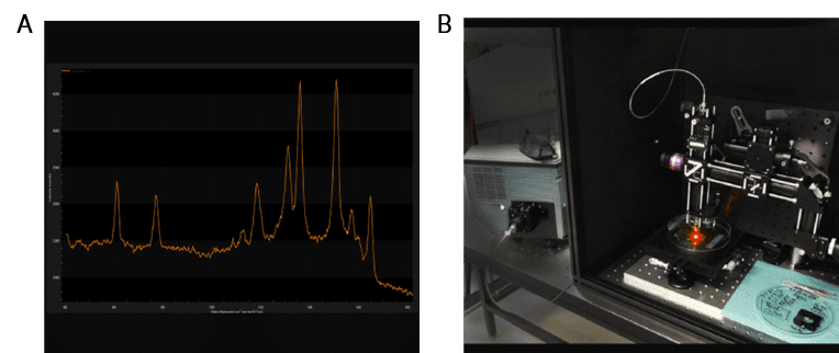
Figure 1: (A) shows a LightField screenshot of a SERS spectrum of cholic acid on a gold nanostar-coated substrate, with data collected using a FERGIE coupled to a laboratory-made confocal Raman microscopy via optical fiber. This experimental set-up can be seen in (B) and is used for the identification of metabolic markers by SERS.
Challenge
One of the challenges that Dr. Boudreau’s research group specialize in is the synthesis of luminescent nanoparticles. These nanoparticles consist of a plasmonic core coated with concentric dielectric layers doped with fluorophores sensitive to various physical and chemical stimuli. As the physical parameters of the particles influence luminescent behaviour, the researchers use single-particle spectroscopy to establish design rules that will result in nanostructures with optimal properties.
Another challenge that the researchers are focusing on is imaging the evolution of key cell metabolites. The same luminescent plasmonic nanomaterials synthesized by the research group are also being grafted on the tip of custom-made optical fibers for remote chemical/biological sensing. This will allow for monitoring of water treatment in plants, and in vivo molecular sensing in model animals.
Dr. Boudreau’s group is also collaborating with other research groups on the development of methodologies based on plasmonic nanomaterials and surface-enhanced Raman spectrometry (SERS) for in situ identification and quantification of cell metabolic markers. Applications include studying the acclimation of phytoplankton to changes in global climate (with Dr. C. Lovejoy, U. Laval, and J.F. Masson, U. Montreal) as well as in vivo monitoring of cholic acid derivatives in the gut microbiota (O. Barbier, A. Marette, and R. Vallée, U. Laval).
The ease with which FERGIE can be coupled to an optical setup, either free-space or via optical fiber, makes this [single-particle fluorescence and scattering data] very easy
Solution
Dr. Boudreau’s research group used the FERGIE (previous version of IsoPlane 81), alongside a custom-made confocal microscope, to collect single-particle fluorescence data and scattering data of the fabricated luminescent plasmonic nanoparticles. This was then correlated with electron microscopy data to fully characterize the nanoparticles. As FERGIE can be coupled to an optical setup, it was easy for the researchers to couple via either free-space or via optical fiber to obtain these measurements.
The high sensitivity and multichannel capability of the FERGIE system allowed Dr. Boudreau’s research group to simultaneously capture any light emitted from multiple species-selective molecular probes on the tip of the fiber, onto which luminescent plasmonic nanomaterials were grafted. As FERGIE is able to switch easily between imaging modalities, the research group found it a useful tool for obtaining multiple measurements from one system, switching between the microscope to a microfluidic flow chamber to the connectorized fiber sensor in mere minutes.