X-ray Imaging
Due to their short wavelengths, x-rays can be used to image samples on the nanoscale. There are multiple different types of x-ray imaging, such as x-ray tomography, or coherent diffraction imaging. Imaging can be combined with microscopy or spectroscopy to find out further information about a sample, such as chemical composition.
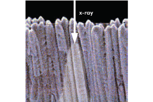
X-ray tomography enables 3D reconstruction of internal structures of real objects non-destructively, all while maintaining a high spatial resolution. By detecting either the attenuation or the phase shift of the transmitted beam at various angles, 2D slices of the microstructure of a material can be reconstructed. These 2D slices can be stacked together to produce a 3D image of the material.
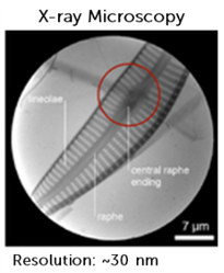
Coherent diffraction imaging (CDI) is a lensless technique which reconstructs an image from the diffraction patterns of a sample. Reconstruction of the image is achieved via an iterative feedback algorithm which retrieves the lost phase information. In this way the lens of an instrument is replaced by the algorithm. CDI is able to provide 2D and 3D information about a material, providing high spatiotemporal resolution for probing dynamic phenomena.
Application Notes
Introduction to X-ray Microscopy
The ability to image smaller and smaller samples allows us to see the previously unseen and analyze even the smallest parts of the universe, from viruses to proteins and even the atomic structure of materials.
There are several different techniques available when performing microscopy, each with its own maximum resolution and imaging capabilities. These include light microscopy, electron microscopy, and x-ray microscopy…Read Full Article
X-ray μCT Provides Nondestructive, High-Resolution 3D Imaging
Since the early 1970s, x-ray computed tomography (CT) has been one of the most versatile, non-invasive investigative techniques in the medical field. It has also enabled nondestructive investigations in many other fields over the past few decades, including industry, archaeology, life science, geoscience, and crime investigations. Unfortunately, conventional x-ray CT systems are only able to achieve spatial resolution at the…Read Full Article
Technical Notes
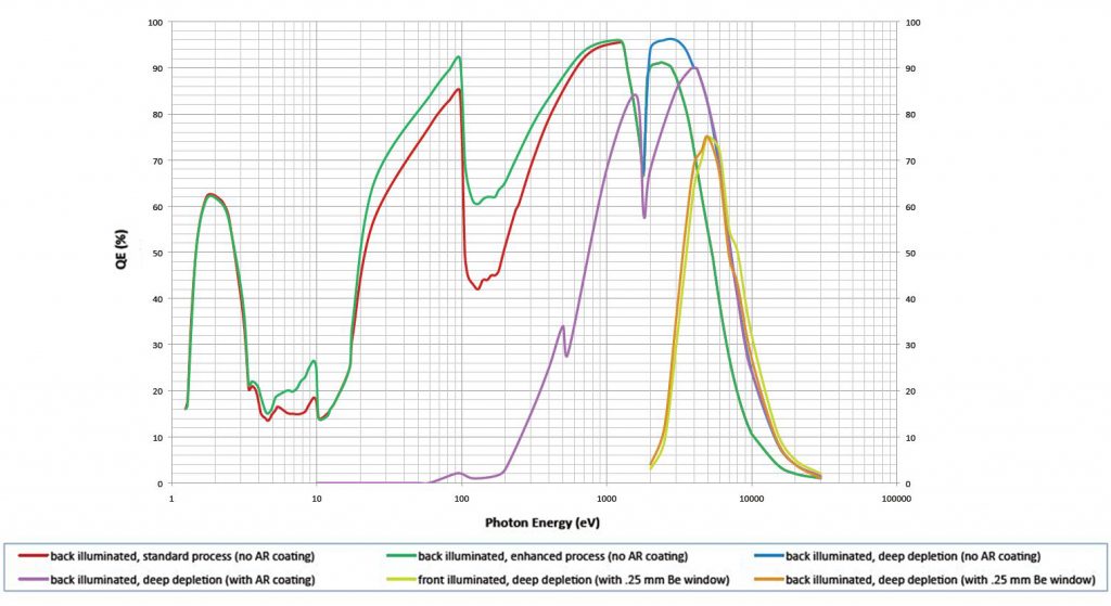
New QE Response Curves for Soft X-ray to VUV Energy Range
Since the invention of charge-coupled-device (CCD) technology in 1969, its extended sensitivity from the NIR to the x-ray region of the electromagnetic spectrum has been utilized to good effect in a wide variety of application areas.
Owing to the unique characteristics of their construction, CCDs are especially useful for imaging and spectroscopy performed in…Read Full Article
Flexible Electronic Architecture Extends Utility of Scientific Cameras
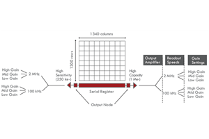
Ultimately, the true worth of a scientific camera is determined by its ability to flexibly meet the performance requirements deemed most useful by a given researcher. As many disciplines have continued to evolve over recent years to encompass more varied investigatory techniques, sets of critical requirements have also expanded. When seeking a scientific camera to satisfy diverse needs, one must be sure to look for the most versatile, highest performance solutions available…Read Full Article
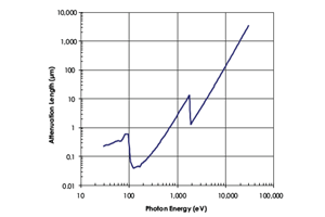
Direct Detection of X-rays (30 eV to 20 keV) Using Detectors Based on CCD Technology
CCDs have become increasingly specialized to meet the changing requirements of both commercial and scientific markets. In the scientific market, CCDs have been improved and optimized in a variety of ways to provide high performance across a broad set of applications — from spectroscopy and semiconductor testing to biological imaging and genetic research…Read Full Article
Utilizing Fiberoptics for Indirect Detection of X-rays
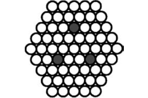
Ultimately, the true worth of a scientific camera is determined by its ability to flexibly meet Quite often, users of indirect-detection systems assume that the camera manufacturer has carefully selected the ideal combination of sensor, fiberoptic (faceplate/taper) and phosphor to preserve image quality. Unfortunately, each application requires different parameters, and it is difficult for a manufacturer to know every requirement. Therefore, it is imperative that each customer understands the component options available to obtain optimal performance.…Read Full Article
Utilizing Phosphors for Indirect Detection of X-rays
Teledyne Princeton Instruments has developed two types of phosphors for x-ray imaging applications in the energy range between 5 keV and 50 keV. The Gd2O2S:Tb is recommended for x-ray energies <33 keV due to its higher absorption efficiency, while the CsI:Tl is recommended where higher resolution is required….Read Full Article