Small Animal Imaging
Small animal research is a key translational link between fundamental research and clinical research. Animal models are utilized for essential preclinical in vivo information, such as sample dosage, for many biomedical therapies and devices prior to clinical acceptance.
When conducting preclinical in vivo studies, there are multiple imaging techniques to gather information. X-ray and UV-VIS-NIR detection has been commonly used within medical imaging, each with their own advantages and limitations. X-rays provide good sample penetration but are unable to give soft tissue information. UV and visible wavelengths can provide soft tissue information but are limited by penetration depth due to the high scattering of these wavelengths within tissue.
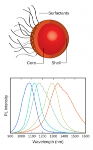
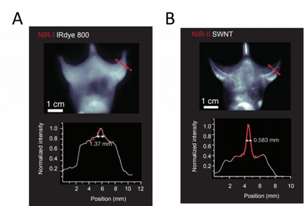
Near infrared (NIR) wavelengths are advantageous as they have reduced scattering within biological soft tissue and are thus used for preclinical in vivo studies. NIR can be separated into two distinct windows, with NIR-I encompassing wavelengths in the 750-1060 nm range, and NIR-II encompassing wavelengths in the 1060-1700 nm range. NIR-I wavelengths can be imaged via silicon CCDs but are only able to penetrate a few millimetres into soft tissue. NIR-II can penetrate deeper into tissue, due to reduced light scattering and autofluorescence, but can only be detected by InGaAs cameras. NIR-II methods have recently been developed for small animal imaging to investigate predominately early disease detection. Majority utilize fluorophores, such as single-walled carbon nanotubes and quantum dots, that fluoresce in the NIR-II region.
Application Notes
NIR-II Probes for In vivo Imaging
Optical fluorescence imaging is one of the most common techniques for imaging in vivo, due to its high temporal and spatial resolution [1]. As it is a non-invasive, real-time technique it is an attractive imaging modality for medical applications such as cancer diagnostics, biosensing, and medical testing.
The majority of fluorescence probes, essential for optical fluorescence imaging, reside within the visible range (400-700 nm). However, visible light is limited when imaging within the body…Read Full Article
Spinning Disc Confocal Microscopy In The NIR-II Window

Near-infrared (NIR) fluorescence is a technique used widely within biological and medical research due to the fact that NIR light can penetrate deeply in biological specimens. It offers high spatiotemporal resolution alongside the capability to image quickly (Fan 2019). The second NIR window (NIR-II, 900 – 1700 nm) is of recent interest due to its superior penetration depth, reduced tissue absorption and…Read Full Article
In vivo Fluorescence Imaging in the NIR-I Spectral Region for Early Cancer Detection
UV, VIS, and NIR-I detection methods have been used in various scientific and medical applications for decades. Each of these approaches, however, has its limitations. For instance, light at UV and visible wavelengths is quite easily detectable using silicon-based CCD technologies but is unable to penetrate samples due to reflection and scattering.
New CCD cameras can detect NIR-I wavelengths from 750 nm up to almost 1100 nm and thus provide…Read Full Article
Deep-Cooled InGaAs FPA Camera Enables High-Speed, High-Resolution In vivo Imaging of SWIR-Emitting Quantum Dots
Working in the shortwave-infrared (SWIR) region of the spectrum affords researchers several advantages, including the abilities to circumvent unwanted fluorescence backgrounds and to probe more deeply into sample surfaces. The advent of deep-cooled camera systems that employ indium gallium arsenide (InGaAs) focal plane arrays (FPAs) has further increased…Read Full Article
Aberration-Free Spectrographs and NIR-Sensitive InGaAs Cameras Facilitate the Development of Carbon Nanotube Optical Sensors for Early Disease Detection
One of the main areas of research at the Memorial Sloan Kettering Cancer Center in New York City is the development of nanoscale sensors to detect cancer at its earliest stages. The research group led by Dr. Daniel Heller uses novel nanomaterials with unique optical properties, making it easier to identify disease biomarkers within the body and thus permit detection before symptoms arise. These nanotechnologies also allow…Read Full Article

Deeply Cooled, Scientific InGaAs Cameras Facilitate NIR-II / SWIR Imaging for Drug Discovery / Small-Animal Research
For decades, x-ray and UV-vis-NIR detection methods have been used in various scientific, military, and medical applications. Although generally employed to good success, these systems nonetheless have some limitations when utilized for such types of work. Effectively leveraging the good sample penetration afforded by x-rays, for example, can prove difficult for…Read Full Article
Technical Notes
Optimizing Detection in Whole Animal In vivo Imaging
Animals studies contribute significantly to our understanding of human disease, and function as an established and essential step in the development of treatments and other therapeutic agents. These studies are conducted in the preclinical phase, preceding drug screening in human clinical trials. The aim of these studies is to determine efficacy, safety, dosing, and toxicity, as well as risk-benefit trades. There are many established, standardized methods implementing animal studies in life science…Read Full Article