
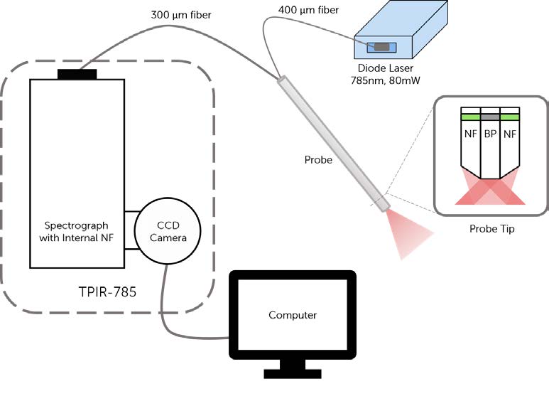
General Raman
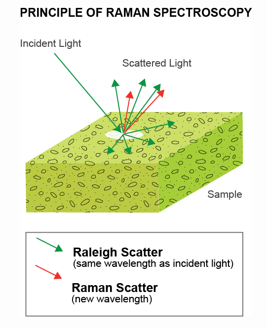
Raman spectroscopy is a non-destructive technique spanning a wide range of scientific and industrial applications. It is often used to characterize or identify the chemical composition and structure of an unknown material. Incident laser light, in the UV, visible or NIR, is scattered inelastically from molecular vibrational modes of the sample.
The frequency difference (measured in relative cm-1) between the incident and scattered photons is called the Raman shift. The majority of inelastically scattered photons are found at positive Raman shifts, corresponding to lower energies and longer wavelengths – this is referred to as Stokes scattering. The scattered photons are analyzed by a spectrometer to produce a Raman spectrum.
With Stokes scattering, the energy of the molecule increases and the Raman scattered photons are red-shifted. With Anti-Stokes, the energy of the molecule decreases – so therefore the molecules must have already been in a vibrationally excited state – and the Raman scattered photons are blue-shifted.
Application Notes
Introduction To Raman Spectroscopy

Raman spectroscopy is an optical scattering technique that is widely used for the identification of materials and the characterization of their properties. It is commonly applied in material science, chemistry, physics, life science and medicine, the pharmaceutical and semiconductor industries, process and quality control and forensics. Raman scattering is an inelastic spectroscopy technique meaning incoming light undergoes a change in color and is scattered with a different energy. The Raman process specifically describes the interaction of incident light with molecular vibrations and rotations in a material….Read Full Article
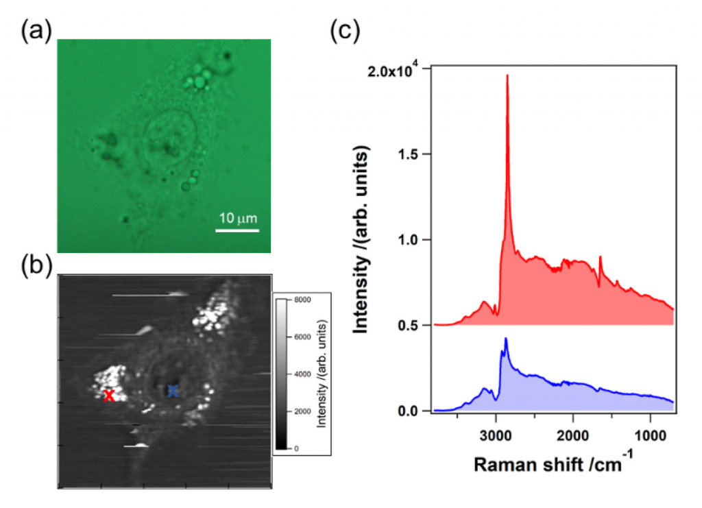
A Brief Overview of Raman Spectroscopy in Life Sciences
Raman spectroscopy has become a widely used tool in biomedical engineering and life science for its diagnostic potential. It is used in medical research, studying the biochemical environment of single cells or monitoring the reaction of cells to drugs, pharmaceutical industry for process and quality control in the manufacturing of drugs. In medical diagnostics Raman spectroscopy has been recognized for its high diagnostic potential…Read Full Article
Deep Depleted CCD Cameras for Raman Spectroscopy In vivo and Medical Diagnostics
Raman spectroscopy is an important measurement technique in life sciences and biotechnology, from nanoscale experiments analyzing the structure of single biochemical molecules to detection of disease and monitoring properties of tissue. Raman spectroscopists in life science research use excitation and detection in all wavelength ranges from the ultraviolet (UV) to the near (NIR) and short-wave infrared (SWIR) region and selection of the laser excitation wavelength is an important experiment parameter to balance spectral resolution, detection efficiency and avoiding autofluorescence background….Read Full Article
Low-Frequency Raman Spectra of Amino Acids: Discovery of a Second Fingerprint Region
A novel astigmatism-free spectrograph design, the Princeton Instruments SCT 320 IsoPlane Schmidt-Czerny-Turner (SCT) spectrograph, is shown to give Raman spectra with better resolution and signal-to-noise ratios than traditional Czerny-Turner (CT) spectrographs. A single-stage SCT spectrograph has been interfaced to a new low-frequency Raman spectroscopy module that…Read Full Article
Using Raman Spectroscopy to Detect Malignant Changes in Tissues
Accurate, rapid and non-invasive detection and diagnosis of malignant disease in tissues is an important goal of biomedical research. Optical methods, such as diffuse reflectance, fluorescence spectroscopy, and Raman spectroscopy, have all been investigated as ways to attain this goal. Diffuse reflectance utilizes…Read Full Article
Technical Notes
A New Dawn for NIR Spectroscopy

Featuring two revolutionary back-illuminated deep-depletion sensors, Teledyne Princeton Instruments BLAZE cameras for spectroscopy provide the highest near-infrared quantum efficiency, fastest spectral rates, and deepest thermoelectric cooling available in a CCD platform. Lower thermally generated dark noise, combined with…Read Full Article
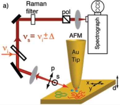
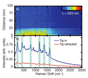
Tip-Enhanced Raman Spectroscopy
TERS works in conjunction with scanning probe microscopy (SPM), a technique that utilizes a metal tip to probe the size and shape of samples at atomic dimensions.
The tip can be coated with silver or gold and, when brought close to a sample of molecules, excited with a laser yielding a spectra with greatly enhanced Raman signal.
TERS confines the signal enhancement to the near-field surrounding the very sharp SPM tip, allowing optical spatial resolution down to ~ 10 nm. The signal can be so strong that single molecules can be detected, ideal for samples that exhibit heterogeneities on the nanoscale.
Application Notes
Tip-Enhanced Raman Scattering (TERS)
The ability to identify small quantities of adsorbed analyte or structural features down to the few-molecule level is a key challenge in nanotechnology. Optical spectroscopy provides an attractive means to achieve such identification via non-invasive implementations and the potential for chemical sensitivity. Raman scattering has emerged as a particularly powerful technique due to its…Read Full Article
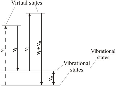
Coherent Anti-Stokes Raman Spectroscopy (CARS)
CARS is a nonlinear Raman spectroscopy technique that uses two very strong collinear lasers to irradiate a sample. The frequency is usually kept constant, with the second laser tuned so that the frequency difference between the two lasers equals the frequency of a Raman-active mode of interest.
To obtain a strong Raman signal, the second laser frequency should be tuned in such a way that its frequency is equivalent to the constant frequency of the first laser minus the frequency of a Raman-active rotational, vibrational, or other mode.
This will cause the frequency of the scattered light to be higher than that of the excitation frequency and so forms anti-Stokes frequency.
Application Notes
Ultra-Multiplex CARS Spectroscopic Imaging of Living Cells
Popular molecular imaging techniques are only able to reveal the distribution or behavior of specific molecules within the human body that have been labeled with pigments or fluorescent proteins. Raman spectroscopy, however, allows researchers to identify the components of…Read Full Article