Small Animal Imaging
Small animal research is a key translational link between fundamental research and clinical research. Animal models are utilized for essential preclinical in vivo information, such as sample dosage, for many biomedical therapies and devices prior to clinical acceptance.
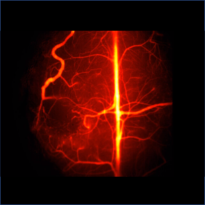
When conducting preclinical in vivo studies, there are multiple imaging techniques to gather information. Optical imaging detection, over the UV to NIR wavelength range, has been commonly used within medical imaging, with each wavelength having their own advantages and limitations. UV and visible wavelengths can provide soft tissue information but are limited by penetration depth due to the high scattering of these wavelengths within tissue.
Near infrared (NIR) wavelengths are advantageous as they have reduced scattering within biological soft tissue and are thus used for preclinical in vivo studies. NIR can be separated into two distinct windows, with NIR-I encompassing wavelengths in the 750-1060 nm range, and NIR-II encompassing wavelengths in the 1060-1700 nm range. NIR-I wavelengths can be imaged via silicon CCDs but are only able to penetrate a few millimetres into soft tissue. NIR-II can penetrate deeper into tissue, due to reduced light scattering and autofluorescence, but can only be detected by InGaAs cameras. NIR-II methods have recently been developed for small animal imaging to investigate predominately early disease detection. Majority utilize fluorophores, such as single-walled carbon nanotubes and quantum dots, that fluoresce in the NIR-II region.
Cameras for Small Animal Imaging
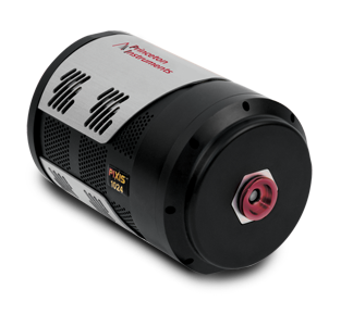
PIXIS
The PIXIS is a low-noise CCD camera with broad sensitivity over the UV to NIR wavelengths (120-1100 nm). This allows for complete experimental flexibility with regards to probe emission wavelength, imaging everything from thick tissue slices to whole live in vivo animal models.
With deep-depleted and eXcelon processed back-illuminated sensor options, the PIXIS is capable of >80% QE within the NIR wavelength range. This is advantageous to in vivo imaging as these longer NIR wavelengths provide deeper penetration depths into samples, while reducing autofluorescence, providing higher quality images than with visible light.
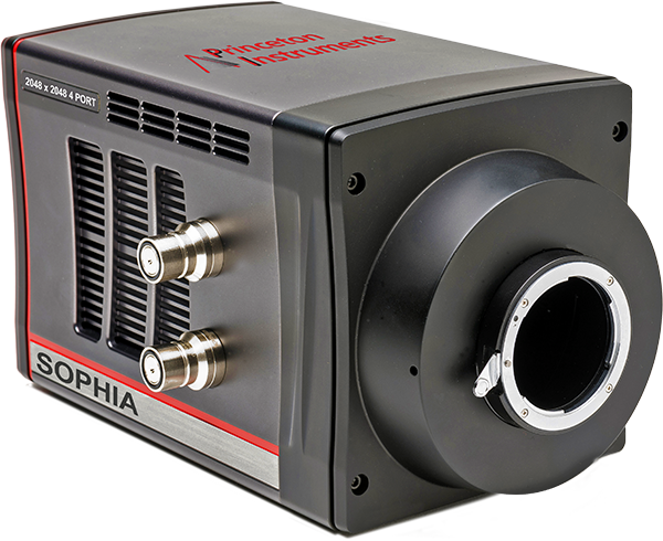
SOPHIA
The SOPHIA camera has a large format CCD sensor for high throughput in vivo imaging over the UV to NIR wavelength range (300 – 1000 nm). The large field of view offers high efficiency, visualizing more of your target tissue while reducing sample photodamage. As more sample area can be imaged, more quantitative analysis can be done on each acquired image.
With deep-cooling ArcTecTM technology, the SOPHIA has ultra-low noise. In addition, the SOPHIA has a high dynamic range and >95% peak QE making it ideal for ultra-low light imaging, common within in vivo studies. The low noise of the SOPHIA makes it perfect for long integration times, providing effective acquisition over a large imaging area.
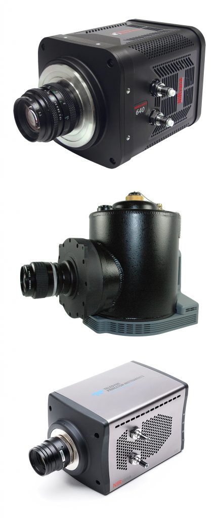
NIRvana
NIR-II fluorescence is used within small animal imaging due to penetration depth and reduced scattering, however a typical silicon based sensor cannot image those wavelengths. The NIRvana family of InGaAs camera features high sensitivity in the 900 – 1700 nm range, ideal for capturing low-light NIR-II/SWIR fluorescence from small animal fluorescence probes.
Noise is reduced with the NIRvana as it is thermoelectrically cooled, with temperatures reaching as low as -85℃, providing the lowest level of dark noise for small animal imaging. The cryogenically cooled NIRvana LN camera is also available, reaching temperatures of -190℃.
With high signal-to-noise ratios up to 250 full frames per second, the NIRvana family allows for fast capture of fluorescence even with extremely low exposure times.
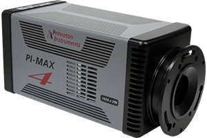
PI-MAX 4
Small animal optical imaging relies on light emitted from molecular probes within the animal. The PI-MAX4 combines the advantages of EMCCDs and ICCDs to deliver excellent precision timing, sensitivity, intelligence and speed for measuring in vivo fluorophores.
The PI-MAX4 offers high quantum efficiency in the wavelength region of 200 – 900 nm, encompassing the NIR-I window.
The coupling of the EMCCD to an image intensifier allows for a 6x higher light throughput between the image intensifier and the detector in comparison to lens-coupled configurations. This ensures imaging of even the faintest fluorophores.