Dr. Joanna Dziadkowiec, Dr. Anja Røyne
University of Oslo
Background
Although we perceive geological processes on macroscopic length scales, the mechanical behavior of geological structures can be significantly influenced by the microscopic mineral structure of rocks as well as the micro-scale interactions at the contacting mineral surfaces. Microscopic and nanoscale spaces between mineral grains often contain fluids and water that can reactively erode or deposit material, e.g. by promoting crystallization processes. Overall, these reactive dissolution and growth processes may have consolidating or destabilizing influences. These can further express in macroscopic, observable changes over time. A better understanding of these processes is important for predicting the behavior of geological structures both by natural development, but also due to human impact, for example, due to large-scale oil drilling or potential capture of carbon dioxide in geological structures.
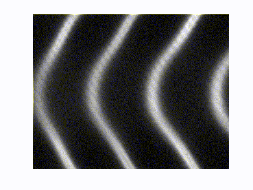
Joanna Dziadkowiec and Anja Røyne performed research at the Njord Center at the University of Oslo trying to understand what happens at contacting mineral surfaces when they are pushed together and exposed to influences of fluids and water. Over time, the solution- and pressure-induced changes in the surface structure alter how the reactive, contacting surfaces interact with each other. This, in turn, affects the overall mechanical behaviour of granular rocks. For their experiments, the researchers use a surface force apparatus (SFA), a measurement device, which determines the interaction force between 2 solid surfaces. Unlike atomic force microscopy, where surfaces are probed point-wise on a nm scale, the SFA technique samples a much larger contact area of up to 100 μm diameter. This scale is more suitable and relevant to relate their observations to geologically relevant processes. For complete characterization of the interaction dynamics, the researchers also measure the distance between the surfaces (imagine a spring where the spring constant is characterized by the applied force and the displacement from the relaxed position). The nm-sized space between the surfaces is measured using multi-beam interferometry where the sample surfaces sit between 2 highly reflective substrates which are illuminated by a white light source. The transmitted signal is imaged onto the entrance slit of a spectrograph and dispersed to visualize fringes of equal chromatic order (FECO fringes). For example, Figure 1 shows the FECO fringes of 2 surfaces in contact with van der Waals forces acting between them. Fringes detected at different horizontal positions in the image correspond to a different wavelength, whereas different vertical positions correspond to different points on the sample surface.
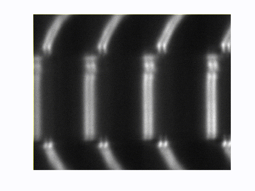
The FECO interference technique allows the researchers to measure the distance profile between the 2 mineral surfaces and detect reactive changes that alter the nano- and micron-scale surface forces and separation distances. By observing the behavior of the interference patterns over time, the researchers are able to investigate reactive changes in the presence of fluids. Figure 2 shows how the interference pattern is modified by the presence of a capillary water bridge between two surfaces placed in close proximity: the capillary bridge is indicated by the discontinuity in the pattern and disappears once water is injected.
Challenge
The spectroscopy system used for multibeam FECO interference in the SFA requires high spectral accuracy and resolution as well as good imaging capability. The changes in the surface profiles of the mineral sample can be in the nm range so a spectral resolution in a nm or sub-nm range is needed to accurately determine fringe positions.
The interference patterns are observed by dispersing the signal along the spectrometer’s entrance slit into a hyperspectral interference image on the system camera, where the horizontal positions, which correspond to wavelength and the vertical position, can be mapped onto different points within the relatively large sample region of interest. The good imaging quality of the spectrograph ensures that the signal is undistorted at all points on the camera. Spectrographs that are optimized for the detection of single spectral channels often show severe image aberrations outside of the focal plane center, leading to distortions, signal reduction, and loss of measurement precision.
“The low aberrations of the IsoPlane system allow us to make very precise measurements of 2D spectral interference patterns. In combination with LightField and Intellical, this system is very efficient to setup for challenging scientific measurements, such as imaging of reactive mineral surfaces in confinement”
Solution
The multi-beam interference setup at the Njord center uses an IsoPlane-SCT320 spectrograph coupled with a PIXIS-2KBUV camera for hyperspectral detection of FECO fringes.
The IsoPlane is an advanced imaging spectrograph with aberration-corrected optics, significantly reducing image aberrations such as astigmatism that could distort the spectral image. The good image quality at any point of the focal plane also increases resolution due to better focusing of the signal light onto the camera. The IsoPlane is ideally suited for multi-channel and hyperspectral measurements requiring this high spectral and spatial resolution.
The PIXIS-2KBUV has a large 2048×512 pixel sensor, covering a wide area in the focal plane, so a wider profile of sample positions can be measured simultaneously. The PIXIS is a deep-cooled scientific camera with very low noise that is optimized for precise, quantitative signal detection.
As the researchers work frequently at different spectral ranges they also use an automated system for accurate wavelength calibration called Intellical. This system, which is controlled by Princeton Instruments LightField software, makes sure that spectral calibration can be done quickly, with very high precision (up to 10x higher than other common calibration methods).
The researchers also have to monitor the interactions between the sample surfaces for long times to be able to detect the reactive changes. Using a time-lapse feature in LightField, the experiments can be easily setup to automatically acquire images for long times with arbitrary time intervals between them, so the acquisition can be optimized considering the speed of the surface evolution.