Prof. John A. Capobianco
Gabi Mandl, Graduate Student
Lanthanide Research Group, Department of Chemistry and Biochemistry, Centre for NanoScience Research, Concordia University, Montreal, Canada
Background
The lanthanide research group of Prof. John A. Capobianco focuses on materials based on the lanthanide group of elements. Originally studying these materials in bulk crystals and glasses, Prof. Capobianco was one of the researchers who pioneered the synthesis and development of nanoparticles containing these elements. The lab is now developing lanthanide nanoparticles with special focus on applications in medicine and biology.
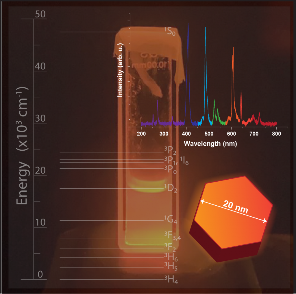
Lanthanide particles have unique optical and luminescence properties and can be tailored chemically to disperse in non-polar solvents as well as biological systems. Lanthanides can produce luminescence through upconversion from longer wavelength and can exhibit a wide emission spectrum ranging from the UV to the IR. In medicine these upconversion processes are relevant for photo activated applications, for example photodynamic therapy for cancer treatment.
PhD candidate Gabi Mandl shared with us insights into her research in the lab focusing on the production of radioluminescent nanoparticles doped with praseodymium. Radioluminescent particles emit light in the UV to visible wavelength range when exposed to ionizing radiation such as X-Rays which makes them viable candidates to consider for radiation therapies.
Spectroscopy plays an important role for characterizing the emission properties and dynamics of lanthanide-based nanoparticles. Different synthesis processes produce various chemical properties that need to be optimized and tailored to specific applications. Specifically, spectroscopy probes the occupation electronic states in the lanthanide ions and how they populate, how fast they emit light, how energy is transferred to other ions, and how the interaction with material defects influences their properties.
Challenge
The lab operates multiple laser systems and high performance monochromators to perform high sensitivity spectroscopy. A CCD based spectrograph can acquire data faster and more efficiently, but should not compromise on sensitivity and spectral resolution. At the same time, the spectroscopy solution should be quickly adaptable to different requirements for various research projects going on in the lab and systems that integrate a CCD and spectrographs were considered for ease of use. Radioluminescence is often much weaker than other forms of luminescence, requiring high detection sensitivity. Measurements of emission dynamics additionally require the ability to operate at high spectral rates and to precisely synchronize the spectral acquisition with external devices. Miniature, integrated spectrographs were considered by the lab due to their ease of use, but they did not provide sufficient detection sensitivity.
“It is mind blowing to me that such a powerful piece of equipment is smaller than the size of a sheet of paper.”
Solution
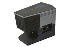
Gabi’s setup for radioluminescence measurements uses a FERGIE spectrograph (now called Isoplane-81) that is receiving light from a fiber optic cable that collects light from samples in a radiation safe chamber. The Isoplane-81 integrates a spectrograph with a high sensitivity, back-illuminated CCD in a compact housing and deep cooling of the camera sensor avoids thermal noise and increases the sensitivity. This system provides great sensitivity for measuring radioluminescence from nanoparticles emitting from 210nm into the visible energy range.
The aberration corrected optical design of the Isoplane-81 system also means that the lanthanide nanoparticle spectra can be measured without compromising spectral resolution, required in Gabi’s experiments, despite the small form factor of the spectrograph. In addition, the system is used by other researchers in the lab by adapting to different fiber and free space optical setups, and fast ability to change entrance slit and grating if resolution requirements change.
Gabi uses LightField data acquisition software to control the spectrograph and allows to customize the user interface with settings required for her experiments so they can be performed more efficiently. To check for the presence of spectral lines or to compare the intensity ratios between different bands LightField provides the ability for quick data visualization by overlaying spectra or quick export to a file format of choice for more in depth analysis.
Gabi mentions that the equipment was incredibly easy to setup and fool proof while delivering the high quality required for her measurements.