Prof. Adi Salomon
Bar-Ilan University, Department of Chemistry
Background
Prof. Adi Salomon’s lab is primarily interested in understanding the interactions of molecules with light at the nanoscale and build devices for sensing molecules using light. The group designs and fabricates metallic nanostructures and uses them to influence light on the nanoscale due to interactions with surface plasmons. These plasmonic devices enhance and focus the light into a deep sub-wavelength volume, depending on their size, shape and arrangement of nano particles as well as the light’s energy. Molecules located in close vicinity to these hotspots can experience strong interactions that changes their physical properties and increases signal response, for example in surface enhanced Raman scattering.
One project in the lab investigates the color and intensity of light transmission through triangular, nano-cavities in metallic surfaces. The color of light can be precisely controlled by changing the distance between just two cavities or by controlling the polarization state of the incoming field (light). The Salomon lab uses different sets of cavities milled in thin metallic films to investigate hybrid modes between molecular transition states and plasmonic modes, to change the interaction strength.
Using plasmonic structures the Salomon lab also performs experiments using plasmonic structures to control second harmonic generation (SHG) of light. SHG is ideal for probing thin layers of molecules on surfaces and as well as molecular events occurring on surfaces. Since inversion symmetry has to be broken for SHG to occur, the observed signal is from the surface of a metal without interference from the bulk of the substrate.
Challenge
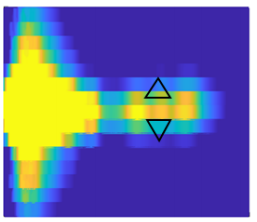
The Salomon lab uses various spectroscopic methods for the characterization of nanostructures such as cathodoluminescence, transmission and reflection spectroscopy. Spectroscopy provides information about the location of electric field hotspots, the position of resonances as well as changes due to changing light polarization and device design. However, it is important to collect this data as a function of position with sufficient spatial resolution. Spectral signals could be obtained by scanning every point on the surface which would be rather slow and inefficient. For this reason, the Salomon lab is using a spectral imaging approach for transmission spectroscopy, where a region of the sample across the plasmonic structures is projected on the spectrometer entrance slit. All spectra from points along the entrance slit will then be measured simultaneously on different rows of the spectrometer camera.
Spectral resolution is also an important consideration particularly when considering molecular spectra. While plasmonic transmission peaks have broad width due to the short lifetime of the surface plasmon excitations, when looking at interactions with molecules fine structure in the molecular spectra contain information about the spectral coupling to the electromagnetic fields.
IsoPlane and PIXIS camera are part of one of the key setups in our lab for characterization of plasmonic structures. The IsoPlane allows us to do spectral imaging without aberrations so hotspots on plasmonic surfaces can be more easily identified.
Solution
The Salomon lab implemented an aberration corrected spectrograph (IsoPlane SCT-320 and PIXIS 1024 camera) in their spectral imaging microscopy setups. This system is designed to minimize or eliminate optical aberrations that are limiting for more conventional Czerny-Turner spectrograph designs. Reducing aberration increases spectral resolution, sensitivity and reduces distortions of sharp spectral lines as the signal is focused on a smaller area in the focal plane. At the same time the aberration free design increases the spatial resolution as spectra coming from different points on the sample are better resolved on the detector. The spatial location and structure of electromagnetic field hotspots in plasmonic structures can be more precisely and rapidly characterized using the spectral imaging approach.