Prof. Yousoo Kim, Dr. Rafael Jaculbia
Surface and Interface Science Laboratory, RIKEN, Japan
Background
The Surface and Interface Science Laboratory at the Japanese RIKEN institute lead by Chief Scientist Yousoo Kim conducts research on surfaces and interfaces at the nanoscale, down to the level of single atoms and molecules. Optical techniques such as Raman spectroscopy are an invaluable tool for investigating these surfaces and new materials in general. However, Raman is impossible to apply to nanoscale materials especially for low analyte counts because of the diffraction limited spatial resolution and the very weak nature of Raman signals.
Rafael Jaculbia, a postdoctoral researcher in the laboratory uses scanning probe microscopy techniques to improve the spatial resolution of Raman spectroscopy. Using a scanning tunnelling microscope (STM), the Raman signals can be enhanced due to the increased electric field at the sharp end of the scanning probes and spatial resolution far below the diffraction limit can be achieved. This technique is called tip enhanced Raman spectroscopy (TERS).
In a recently reported study the lab showed STM-TERS down to 1 nm spatial resolution and single molecule sensitivity. The molecule is placed over a metal substrate to achieve further Raman enhancement through dynamic charge transfer, but is kept in a pristine state by placing the molecule on a few atomic layers of thin salt to avoid the hybridization of the electronic states of the metal substrate and the molecule.
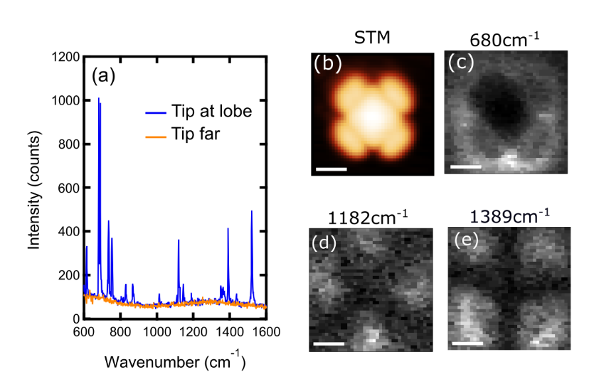
Figure 1 : STM-TERS spectra of a single CuNc molecule comparison with a spectrum several nm away shows that the origin of the Raman signal is localized to the molecule. The images of TERS mapping of the molecule reveal the structure of different vibrational modes.
Challenge
Despite the signal enhancement, Raman signals are inherently weak, which is one of the challenges of single molecule STM-TERS experiments. The spectroscopy system needs to sensitive enough to detect the signals and keeping the measurement time as short as possible for Raman mapping experiments.
Another challenge is the small width (< 5 cm-1) of the spectral lines in the STM-TERS spectra requiring high enough resolution of the spectrograph while keeping high enough light throughput to not lose signal.
We like the high throughput and the good background reduction of the Isoplane. We can highly recommend such a system to those working in in all kinds of Raman spectroscopy such as TERS, resonance Raman, low frequency Raman, etc.
Solution
For the spectroscopic detection for their measurements the lab uses a PIXIS-100BR CCD camera coupled to an IsoPlane-320 Schmidt-Czerny Turner spectrograph. The back-illuminated and deep depleted Silicon sensor of the camera allows for very sensitive detection in the NIR wavelength with up to 95% quantum efficiency. The PIXIS camera sensor is thermoelectrically cooled to -80⁰C or below to reduce thermal excitation and dark current so even low light levels that require minutes of integration time can be detected.
The IsoPlane-320 spectrograph has an advanced optical design that increases throughput, resolution, and sensitivity of STM-TERS measurements. The IsoPlane optical system reduces optical aberrations so incoming photons are focused on less camera pixels resulting in less instrument broadening of spectral lines.
Also, with photons spread out over less camera pixels not only the signal is increased, but also less dark current is detected so measurements will show higher signal to noise ratios. In addition, the increased input aperture allows for high signal collection from the experiment setup.
In their recent measurements Dr. Jaculbia looked at single molecules of copper naphthalocyanine (CuNc)and performed resonant STM-TERS as well as TERS mapping of the molecule with sub-nanometer resolution. Under resonance conditions the STM-TERS mapping reveals the vibrational symmetry of the molecule as evidenced in the spectacular images of different vibrational modes with different energies.
The high resolution STM-TERS technique can be useful in the discovery of new materials and new function of materials. The RIKEN lab will be using the system in the future for further measurements of single molecules as well as 2D materials 2D materials.