Professor Fan Zhang
Dr. Zhang Hongxin, Associate Researcher
Fudan University, China
Background
Prof. Fan Zhang’s research group, from Fudan University, is centred around in vivo fluorescence and biomedical imaging that cannot be detected using traditional fluorophores emitting over the visible wavelength range. His team focuses on creating and characterizing fluorescent probes to detect human disease, as well as imaging approaches to optimize fluorescent probe detection. They test the in vivo ability of these fluorescent probes via animal models, as they are a key translational link between fundamental research and clinical research.
His researchers use both small organic dyes and nanocrystals for in vivo photoluminescence biomedical imaging, as these probes provide fast feedback and high sensitivity. However, all in vivo imaging is limited by tissue penetration depth and image spatial resolution.
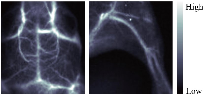
Figure 1: High spatial resolution and SBR imaging of the brain (left) and hind-limb (right) vessels of a mouse obtained based on fluorescent probes with the fluorescence wavelength beyond 1500 nm.
Challenge
To try and overcome this challenge, researchers within Prof. Zhang’s group are creating fluorescent probes that emit in the NIR-II wavelength region (1000-1700 nm). These wavelengths are able to penetrate deeper into the tissue, with minimal light scattering, to provide images with higher spatial resolution.
The researchers are currently using these NIR-II fluorescent probes to evaluate the effectiveness of antihypertensive drugs. They use a murine animal model, imaged within a light restricted system, to determine how the drug is influencing blood vessel structure within the bladder. By using the NIR-II fluorescent probes, they can obtain high resolution images of the blood vessels deep within the tissue, determining any structural changes caused by the drug.
The in vivo NIR-II fluorescence light restricted system, used to evaluate drug efficacy on bladder blood vessel structure, was created by Prof. Fan Zhang’s group, and has now been commercialized at Nuohai life Science (Shanghai) co., Ltd.
The NIRvana 640 is highly sensitive, with a high signal-to-noise ratio, providing clear structural information about the blood vessels
Solution
Prof. Zhang’s research group used the NIRvana 640 InGaAs camera, alongside a custom-built in vivo system, to detect in vivo signals from the NIR-II fluorescent probes. This allowed the researchers to determine any changes in vasculature structure, correlating to the effectiveness of the drug being tested. It also allowed the researchers to characterize the signal intensity of the NIR-II fluorescent probes created in the lab. The longest wavelength emitted by the fabricated fluorescent probes is 1530 nm, so therefore an InGaAs detector was required.
The high sensitivity and high signal-to-noise of the NIRvana 640 allowed clear detection of NIR-II fluorescent probes, providing the blood vessel structural information essential for determining drug efficacy. The quick cooling and high frame rate of NIRvana 640 also allowed the researchers to image very fast and dynamic changes within the animal model.