
Introduction
As is the case with many scientific and commercial technologies, the x-ray imaging and spectroscopy instruments utilized to perform leading-edge academic and industrial research are becoming smaller, more cost effective, and — in a sense — more personalized. For years, researchers across a wide range of disciplines whose daily work relies upon soft x-ray energies have had to schedule sessions at large, high-brilliance x-ray sources in order to conduct critical experiments.1 Time at these expensive-to-operate shared resources (e.g., third- and fourth-generation synchrotrons and x-ray free-electron lasers) remains a precious commodity.
The advent of high-harmonic generation (HHG) instruments, however, is beginning to reshape the landscape for researchers desirous of performing various imaging and spectroscopy experiments in the soft x-ray energy regime. HHG light sources complement large-scale synchrotrons and x-ray free-electron laser (XFEL) sources; among the key advantages of using tabletop HHG light sources for x-ray applications are easier access, full synchonizability to the sub-femtosecond level, and attosecond temporal resolution.2,3
Advanced electronics, nanoscience, semiconductor, materials science, polymer science, biotech, and life science applications can benefit from pairing tabletop HHG light sources with x-ray imaging/spectroscopy detectors to facilitate experimental techniques such as coherent x-ray diffraction imaging (i.e., lensless x-ray imaging), x-ray absorption fine-structure (XAFS) spectroscopy, near-edge x-ray absorption fine-structure (NEXAFS) spectroscopy, and extreme ultraviolet (EUV) spectroscopy.2–7
This application note will address recent work performed by researchers in the Attoscience and Ultrafast Optics group of Prof. Dr. Jens Biegert at the Institute of Photonic Sciences (ICFO) who developed/utilized a tabletop attosecond HHG light source in concert with a Princeton Instruments x-ray CCD camera to demonstrate NEXAFS spectroscopy at the socalled water window’s carbon K-edge of a polyimide foil and retrieve the specific absorption features corresponding to the binding orbitals of the carbon atoms in the foil.4,5
Experimental Setup
The K-absorption edges of carbon, nitrogen, and oxygen occur within what is often referred to as the water window, a range of wavelengths (2.3 nm – 4.5 nm) at which water is virtually transparent. Because each of these elements is fundamental to life, the ability to make increasingly more sophisticated observations in the water window is critical to leading-edge research being conducted by biologists, chemists, and physicists across many diverse fields.5
Until recently, such observations relied on the utilization of large-scale, high-brilliance sources. The aforementioned research group from ICFO in Barcelona is among a number of groups around the world2,6 who are implementing tabletop HHG instrumentation as a compact, cost-effective alternative.
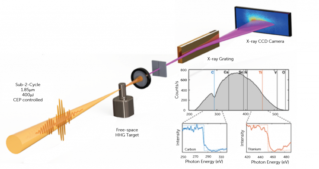
The ICFO group, for example, has developed a tabletop HHG light source to perform high-resolution x-ray absorption spectroscopy on a polyimide foil. Their source rests on a highly stable kilohertz laser system that drives an optical parametric amplifier with subsequent hollow-core-fiber pulse compression, and results in sub-2-cycle carrier-envelope-phase–stable (CEP-stable) laser pulses with a central wavelength of 1.85 μm; the high harmonics generated by this source have reached photon energies up to 535 eV, far beyond the carbon K-edge.4,5
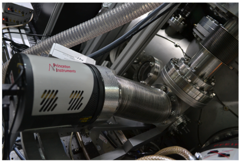
The group used second-harmonic-generation frequency-resolved optical gating (FROG) to measure compressed pulses, and achieved bright soft x-ray flux and a 535 eV cutoff in the water window by focusing the CEP-stable sub-2-cycle pulses with an f = 100 mm silver-coated curved mirror into the HHG target. The harmonic spectra were resolved with an x-ray spectrograph and a cooled Princeton Instruments PIXIS-XO x-ray CCD camera.4 Refer to Figures 1 and 2. The researchers measured the resolution of their x-ray spectrometer as 0.25 eV at 300 eV.
Results
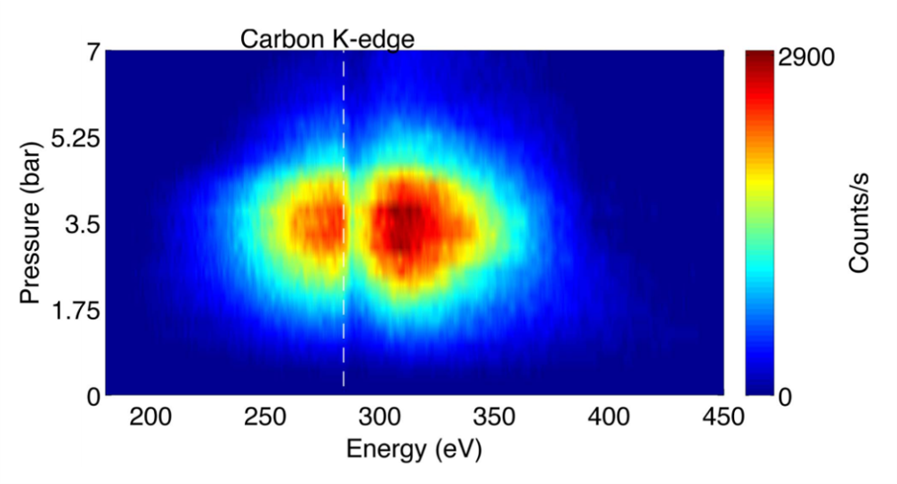
The researchers at ICFO optimized harmonic flux by scanning the target-backing pressure from 0 to 7 bar of neon. Figure 3 shows the results of integrating the HHG spectrum (for a given backing pressure) for 5 sec in steps of 0.25 bar per spectrum; it clearly illustrates that a cutoff far beyond the 284 eV carbon K-edge was achieved and that the highest yield was reached (together with the highest cutoff) at a backing pressure of 3.5 bar.4
After determining the optimum pressure for the target, CEP control of the spectral shape was investigated by measuring x-ray beam brilliance and photon flux. This control was demonstrated to be crucial for avoiding potential over-averaging of pre- and post-edge structures that vary from shot to shot while integrating a NEXAFS spectrum.4
The photon flux of the HHG light source is the highest reported in the water window and, more important, it corresponds to the first isolated 355 attosecond duration soft x-ray pulse. Critical for absorption spectroscopy is the spectral stability, which is ensured through CEP stability of the laser source.3,4 The ICFO research group then demonstrated the utility of their tabletop HHG light source for performing carbon K-edge NEXAFS spectroscopy on a 200 nm freestanding polyimide film. Figure 4 shows the absorption spectrum that was taken in a mere 5 min with all peaks around the carbon K-edge clearly visible and identifiable from the known orbitals in polyimide.4
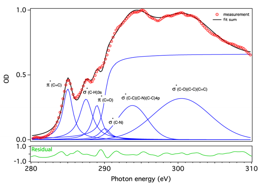
This HHG instrument is the first reported high-flux tabletop source of coherent x-ray radiation in the water window that reaches a photon flux of (1.85 ± 0.12) x 107 photons/sec/1% bandwidth at 300 eV and the first isolated attosecond pulse in that regime.3,4
Enabling Technologies
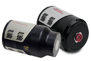
The Princeton Instruments PIXIS-XO scientific camera utilized by the research team in Barcelona features a cooled, back-illuminated CCD without anti-reflective coating in order to facilitate the direct detection of ultra-low-energy x-rays. In addition to the camera’s software-selectable gains and readout speeds, a rotatable conflat flange with a high-vacuum-interface design makes the PIXIS-XO an excellent choice for ultra-high-vacuum applications.
Princeton Instruments offers a variety of cameras for the direct detection of soft x-rays (see Figure 5), including not only the PIXIS-XO but the PIXIS-XB, which uses a thin beryllium window to vacuum seal the unit for deep cooling, protect its back-illuminated deep-depletion CCD, and reduce background by filtering low-energy x-rays.
Another Princeton Instruments scientific x-ray CCD camera, the PI-MTE, implements a thermoelectrically cooled design that utilizes PCBs thermally linked to circulating coolant in order to provide reliable operation inside vacuum chambers. The PI-MTE camera’s compact size and flexible tubing permit the positioning of the detector in limited space or on a movable arm.

Future Trends
The successful use of a tabletop HHG instrument by researchers at ICFO to perform carbon K-edge NEXAFS spectroscopy within the water window embodies the trend towards miniaturization and personalization of coherent x-ray radiation sources for advanced x-ray techniques. The group’s most recently published HHG-enabled results, the spatio-temporal isolation of attosecond soft x-ray pulses in the water window,3 signifies yet another important stride on this march of technological progress.
Researchers from the Far East6 to the Far West2 are designing and utilizing a variety of tabletop HHG light sources to meet their own specific x-ray imaging and spectroscopy requirements. In addition to the obvious cost and access advantages over large-scale, state-of-the-art synchrotrons and XFEL sources, these compact HHG instruments are beginning to offer superior temporal resolution.3 Furthermore, HHG-enabled coherent x-ray diffraction imaging is able to produce quantitative, high-contrast phase and amplitude images in both transmission and reflection, as well as probe dynamic phenomena in three dimensions,2 offering a powerful complement to surface-scanning technologies such as atomic force microscopy.
Scientific x-ray CCD cameras, such as Princeton Instruments’ PIXIS-XO, PIXIS-XB, and PI-MTE, ensure the sensitivity, speed, and flexibility needed to match ongoing advances in tabletop HHG instrumentation. And as x-ray experimental setups for individual labs continue to evolve and gain popularity, selecting the right application-appropriate camera will be imperative to capitalize fully on the benefits of these new setups.
References
- Coherent x-ray diffraction imaging. Princeton Instruments Application Note (2009).
- Adams D.E., Wood C.S., Murnane M.M., and Kapteyn H.C. Tabletop high harmonics illuminate the nano-world. LFW, 38–41 (2015).
- Silva F., Teichmann S., Cousin S.L., Hemmer M., and Biegert J. Spatiotemporal isolation of attosecond soft x-ray pulses in the water window. Nat. Commun. 6 (2015).
- Cousin S.L., Silva F., Teichmann S., Hemmer M., Buades B., and Biegert J. High-flux tabletop soft x-ray source driven by sub-2-cycle, CEP stable, 1.85-μm 1-kHz pulses for carbon K-edge spectroscopy. Opt. Lett. 39, 5383–5386 (2014).
- Cousin S.L., Silva F., Teichmann S., Hemmer M., and Biegert J. Molecular fine structure from water window x-rays. OPN, 58 (2014).
- Sung H., Kim Y.S., Cho W.J., Kim Y.T., Kim J.H., Lee S., and Jhon Y.M. Coherent EUV light source by high harmonic generation for EUV metrology. Recent Advances in Telecommunications and Circuits, 34–36, World Scientific and Engineering Academy and Society Press (2013). ISBN: 978-960-474-308-7
- Coherent diffraction microscopy and its applications in nanoscience and biology. (accessed online Aug. 2015) http://www.physics.ucla.edu/research/imaging/Research/Research_1.pdf
Further Information
Related Products
Overview of the advantages of the products available which are optimized for soft x-ray microscopy applications.

Get In Touch
Speak to a member of our team about your specific application, and what we can do to help you.

Join Our Mailing List
Want to be up to date on the latest offers and news from Teledyne Princeton Instruments? Join our mailing list today.