Wei-Chuan Shih
arXiv
Plasmonic nano-aperture label-free imaging
Introduction
A novel imaging technique using plasmonics enables observations of nanoparticles with enhanced sensitivity.
Prof. Wei-Chuan Shih and his coworkers from the Nano-bio Photonics lab at the University of Houston are researching the use of nanomaterials and devices in applications related to biomedicine, energy and the environment. Using plasmonics the group developed a new imaging technique based on localized surface plasmon imaging that can detect particles with a smaller diameter than 25 nm.
The researchers refer to the technique as PANORAMA (ultra near-field modulated plasmonic nano-aperture label-free imaging). Compared to other plasmonics based imaging techniques, PANORAMA achieves very high spatial and vertical resolution using localization and evanescent wave properties of localized plasmon effects. The technique also uses ordinary bright-field illumination to produce larger signal strength that can be used for fast imaging of nanoparticles on mm-exposure times.
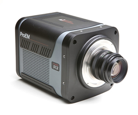
The experimental setup uses an inverted microscope with a ProEM 1024 EMCCD camera for detection where a 20 nm wide bandpass filter is placed in the Fourier plane of a 4f optical system that is located between the sample and the camera. The filter is chosen to be on the red side of the extinction due to localized surface plasmon resonance.
The sample is illuminated through an array of Au nano disks with average size 360 nm and 50 nm thickness. If an object of interest enters the nearfield range of the disk array, the changes in refractive index cause a redshift of the extinction and increased light transmission that can be detected on the camera.
The group demonstrates that the technique can be used to compare the size of polystyrene nanoparticles to sizes below 25 nm as well as to measure the dynamics of particles approaching the surface. Due to the bright illumination, good signal to noise is achieved even for short exposure times in the millisecond range. Moreover, the technique does not suffer from effects causing bleaching or low emission as no fluorescent labels are being used.
In the future the technique could be combined with surface functionalization methods to investigate nanoparticles, vesicles and viruses in biomedical sensing and imaging applications.