Quan Liu
The Journal of Physical Chemistry Letters
Depth-Sensitive Raman Spectroscopy for Skin Wound Evaluation in Rodents
Introduction
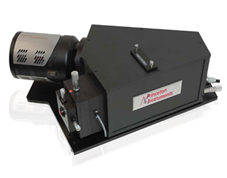
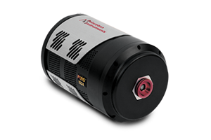
Researchers at Nanyang Technological University in collaboration with others demonstrate design/optimization of a new snapshot depth sensitive Raman instrument for skin sample measurements. Their setup uses the Acton LS785 Spectrograph and PIXIS camera for Raman spectroscopy. Their depth sensitive measurement performance is characterized in both an ex vivo porcine skin with a gradient of layer thickness and an ex vivo rat skin sample with surface wounds. The exposure time for their experiment was well below the permissible exposure as per American national standards.
By using a signal collection fiber bundle, the researchers were able to adjust for variable depth profiles on the sample. They demonstrate simultaneous data acquisition at five different target depths of the sample. This correlates to very good efficiency for an in vivo scenario. PLS-LDA analysis created classifiers between the wound edge and healthy skin based on the acquired spectra. The instrument can discriminate between the wound edge and healthy skin with good accuracy which paves the way towards in vivo preclinical studies of rat skin wounds.