Hiromichi Kataura
Scientific Reports
Introduction
Hiromichi Kataura from the National Institute of Advanced Science and Technology in Japan is well known in the scientific community for his research on physics and applications of carbon nanotubes. Recently him and his team are using carbon nanotubes as small bio probes to image physiological and biochemical activity in-vivo.
Specifically, the researchers are looking at brown adipose tissue (BAT). BAT plays an important role in the regulation of body temperature and when stimulated, anti-obesity and diabetic effects are increased. A deep understanding of the mechanisms in BAT is important for the development of drugs to fight metabolic syndrome.
The researchers describe that the current standard method for observing BAT is positron emission tomography (PET) or histological investigation (ex-vivo). While PET is useful it does not always reflect the metabolic activity of the fat tissue well, so one of Kataura’s goals is to provide new observation methods that correctly correlate to metabolism and allow for in-vivo observations.
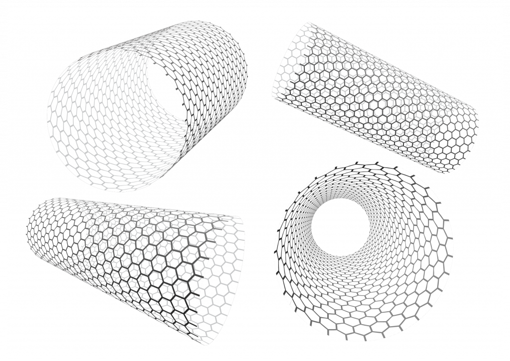
The team prepares carbon nanotubes emitting fluorescence light between 1000-1400 nm where light can penetrate deeply in tissue without absorption or scattering by biomolecules. This range also has low tissue autofluorescence. The nanotubes are can be highly adapted to be specific to the BAT tissue. The team shows that the fluorescence signal can be successfully applied to observe the activity in BAT tissue.