Brian Pogue
Journal of Biomedical Optics
Cherenkov excited short-wavelength infrared fluorescence imaging in vivo
with external beam radiation
Introduction
Radiation therapy is one standard method for treating certain types of cancerous tumors. The challenge in all radiation therapies is always to precisely localize the diseased tissue and deliver a lethal radiation dose at exactly that location without damaging surrounding healthy tissues. Brian Pogues group from Dartmouth in Vermont has pioneered the use of gated ICCD cameras to detect Cherenkov light that gets produced by ionizing radiation in matter. Measuring the Cherenkov emission allows this group to measure radiation deposited at certain locations within a human body.
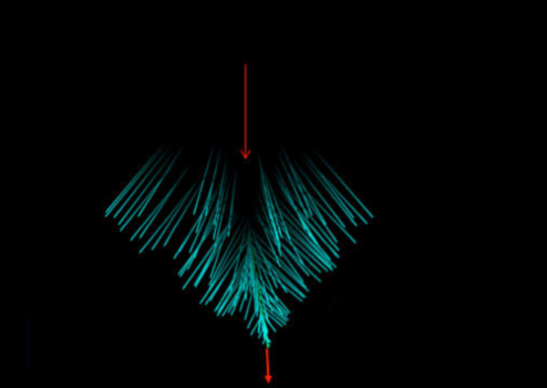
This technology is currently commercialized by a company spun off from Prof. Pogues group. While Cherenkov ligh is mostly emitted in the blue to visible region, SWIR light gives much higher accuracy about the position of radiation in tissue due to the low scattering and absorption by tissue. The researchers are now reporting on using fluorescent quantum dots that are distributed in a tissue sample and designed to absorb the Cherenkov radiation and emit SWIR radiation. The group is now working on applying this technique in radiation therapy as well as imaging molecular processes with targeted nanoscale probes.