Linlin Zhou
Micromachines
Four-Dimensional Stimuli-Responsive Hydrogels Micro-Structured via Femtosecond Laser Additive Manufacturing
Fluorescence imaging helps to characterize hydrogels manufactured for in human tissue.
There is a growing need for micro- or nanostructures machines that mimic dynamic human tissues for applications such as drug delivery, tissue engineering, and nanomechanics. Although this area of research has previously been focused on 3D-printed constructs, they are limited by their bulky size and rigidity. Hydrogels, however, are an emerging alternative and have shown promise in many applications such as cancer treatment and wound healing as they act similarly to human tissue. In addition, hydrogels have good biocompatibility and degradability, with the ability for cell adhesion and growth.
To fully utilize hydrogels for these applications, controllable stimuli-responsive behavior is essential. Additive manufacturing methods have been shown to produce stimuli-responsive behavior in a controlled way. In addition, additive manufacturing allows for a 4D construct, which takes advantage of time-dependent behavior of stimulus-responsive materials such as hydrogels.
Researchers at Jiangsu University, Huazhong University of Science and Technology, and Johns Hopkins University used two-photon polymerization, a typical additive manufacturing method, with a femtosecond laser to create hierarchical microstructures and nanostructures. These structures created a controllable surface tension mismatch. They were able to show that hydrogels with this controlled stimuli-responsive behavior were able to focus a bright spot of light by swelling and shrinking, morph in shape in a similar manner to how plants move, and deform by absorbing localized light energy.
Fluorescence imaging was one method of nanostructure characterization used by the researchers. Fluorescence images were used to determine the structures of the fabricated hydrogels. The IsoPlane and KURO were also used to image the biocompatibility of the hydrogels. To test this, active cells were introduced to the hydrogels, and fluorescence images indicated that the active cells crawled onto the prepared surface, indicating desirable adhesion and biocompatibility for cells.
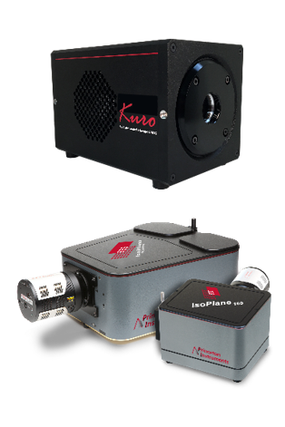
The reflected fluorescence images of the hydrogel structure were captured using an IsoPlane-320 spectrometer and KURO sCMOS camera. The IsoPlane-320 is ideally suited for sensitive micro- and imaging spectroscopy measurements. The advanced optical design of this spectrometer make it possible to achieve higher spatial localization of the measurement signal and reliable measure spectra of several points simultaneously with clear separation of spectral signals. The IsoPlane is also able to capture broadband as well as high resolution spectra with a lot of spectral details. The KURO sCMOS camera features a scientific, back-illuminated CMOS sensors. The high quantum efficiency of the sensor enables measurement of weaker signals with higher S/N ratio. The low readnoise of the camera is ideal for applications where many spectral images have to be read out with high measurement rates.