Zhu Zhu
Acta Biomaterialia
Construction of adipose tissue using a silica expander capsule and cell sheet-assembled of decellularized adipose tissue
NIR-II in vivo and in vitro imaging enable detailed observation of blood vessel growth in enhanced adipose tissue scaffolds.
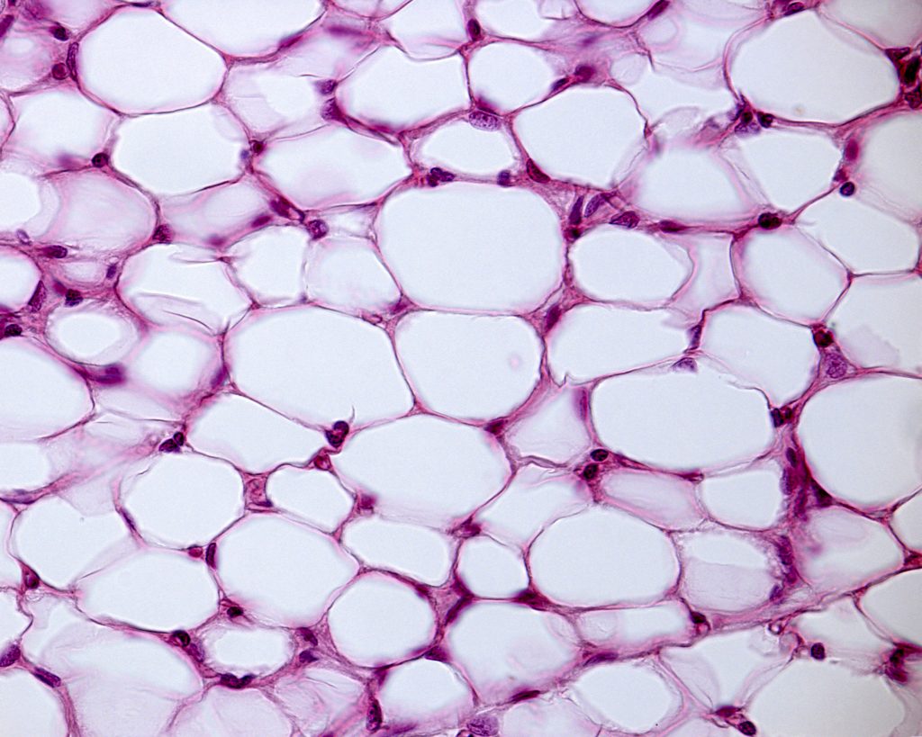
Defects in soft tissue, often caused by burns, congenital deformities, and tumor resections, have long been a common difficulty within reconstructive surgery. Engineered, vascularized adipose tissue has been extensively studied as a potential aid for repair of soft tissue damage or defects. Typically, when utilizing adipose tissue scaffolds, adipose-derive stem cells are introduced to promote tissue regeneration and healing of the soft tissue defect.
However, adipose tissue scaffolds are limited as typically they display delayed neovascularization, the process in which new blood vessels are formed, and unstable adipose formation. Researchers from China have begun tackling these limitations by introducing CCL2, an exogenous chemokine,which enhances both adipose tissue and blood vessel growth. To counteract the effects of delayed vascularization, the researchers also introduced a vascularized fibrous capsule to promote neovascularization.
The researchers studied the effects of CCL2 alone and CCL2 and the capsule in both in vitro and in vivo environments. In order to visualise neovascuarlization in vivo, the researchers obtained NIR-II fluorescence images with a NIRvana camera. They used PbS QDs which were administered immediately before images were acquired. These QDs are advantageous as they have very good biocompatibility and fluoresce well within the NIR-II window, at approximately 1300 nm. This wavelength is beneficial for in vivo imaging as it allows for deeper penetration depth with less scattering and lower tissue autofluorescence, resulting in better image quality.
However, these wavelengths are unable to be imaged using traditional silicon cameras, such as CCD or sCMOS. Instead InGaAs cameras, such as the NIRvana camera, must be used. In addition, the NIRvana InGaAs camera has deep cooling and ultra-low dark current, optimized for long acquisition times typical in in vivo imaging. Furthermore, the high quantum efficiency of the NIRvana means it has high infrared sensitivity for faint signals common when imaging in vivo blood vessels. The researchers found that the use of CCL2 and the capsule provided the highest percentage of blood vessels, with strong indication that new blood vessels had developed.