Sungho Jeong
Journal of Biomedical Optics
Mapping of cutaneous melanoma by femtosecond laser-induced breakdown
spectroscopy
Introduction
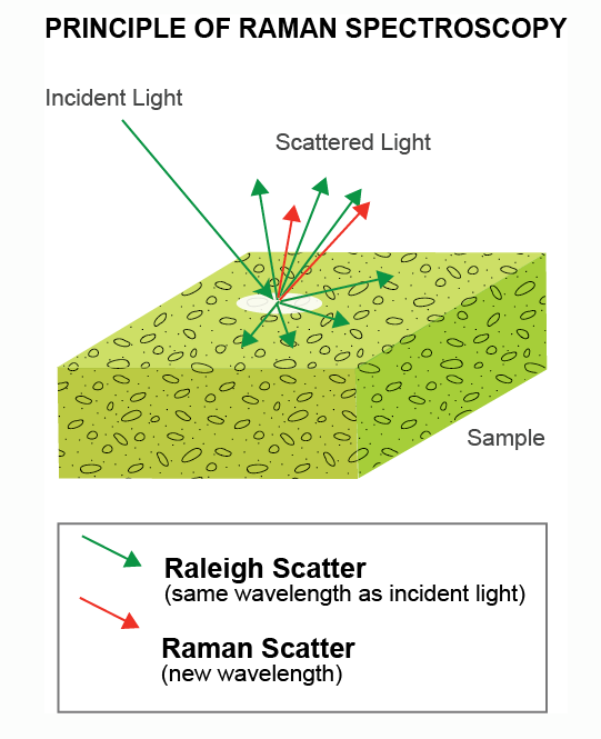
Various optical and spectroscopy techniques are known to help with imaging and identification of cancerous tissue. For example, during surgical removal of cancerous tissue, optical methods like Raman spectroscopy promise to deliver diagnostics about the success of the surgery, such as no remaining cancerous tissue, within seconds to minutes rather than the long wait times associated with conventional lab-based methods.
Besides Raman spectroscopy other optical methods can be used for cancer identification. Researchers around Sungho Jeong from the Gwangju Institute of science and technology are using femtosecond laser induced breakdown spectroscopy (fs-LIBS) to investigate melanoma – one of the most dangerous forms of skin cancer. The rapid and high spatial resolution of the fs-LIBS method allows them to create high resolution allows them to create precise maps of the boundary between cancer and normal skin after surgical removal. The researchers found that certain element signatures like peaks from carbon are basically unaffected between the different skin tissues. However, Mg and Ca elemental peaks show significant intensity changes and can be used as biomarkers to identify cancerous tissue.
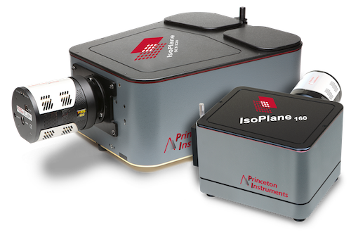
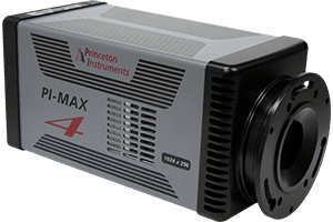
The researchers chose femtosecond laser pulses over longer nanosecond laser pulses as they can achieve higher resolution, affect a smaller tissue region and produce lower background emission. For detection a fast ICCD coupled to a high-resolution spectrograph is used for precise detection of the signal over the background radiation. The team believes that this technique can become a good option for more rapid diagnostics of surgical removal procedures and should be able to be applied to other types of cancer as well.