Tara Sabo-Attwood
NanoImpact
Introduction
Several analytical methods have been commonly employed for carbon nanomaterial detection including electron microscopy (TEM/SEM), Raman spectroscopy, and mass spectrometry. However, each has limitations, for example a lack of spatial resolution specificity for differentiating carbon nanomaterials from the carbon in the biomass of living organisms. Near IR fluorescence imaging has emerged as promising tool for detection and quantification of singlewalled carbon nanotubes (SWCNTs) for in vivo studies.
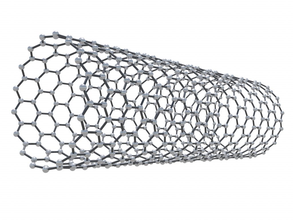
Collaborating researchers from the University of Florida, University of Texas and Duke University in the US published a study demonstrating that near infrared fluorescence was an effective method for pulmonary retention of SWCNTs in a murine model. They used a custom imaging system based on detection with a Princeton Instruments OMA V, 2D InGaAs array for capturing the emission from carbon nanotubes. The concentration of nanotubes is deliberately chosen to be at or below the detection threshold of other technologies such as Raman spectroscopy. The experiments use pristine, unaltered and not functionalized nanotubes. The methods are also applied on tracking and quantifying concentration of carbon nanotubes in lung tissues to assess pulmonary retention and distribution leading to potential health effects in mice.