Cynthia McCollough
European Radiology
Improved coronary calcification quantification using photon-counting-detector CT: an ex vivo study in cadaveric specimens
Introduction
Cardiovascular disease is one of the most common causes of death worldwide. One of the typical indicators of this disease is calcifications within the coronary artery. The most established method for measuring these calcifications is computed tomography (CT). Within the clinic, these CT scanners rely on energy-integrating detectors, which utilize a scintillator to convert high energy photons into visible light. However, they are limited in spatial resolution which can result in either an overestimated diagnosis or an underestimated one. In comparison, photon-counting detectors, currently not used in clinic, use a semiconductor to convert x-ray photons to electrical signals, allowing for smaller signals and improved spatial resolution.
Researchers from Linköping University and the Mayo Clinic in Rochester (USA) have compared a photon-counting detector CT with an energy-integrating detector CT to determine which method is more accurate at quantifying calcifications on the coronary artery. They relied on micro-CT measurements as the reference standard.

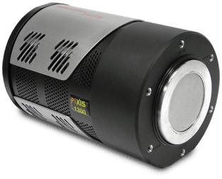
These researchers found that the photon-counting detector CT was not only able to quantify coronary calcifications much more accurately, but also produce lower noise than the standard energy-integrating detectors. The researchers utilized a PIXIS-XB within their micro-CT set up, creating a custom-built micro-CT scanner to create the standard reference. By accumulating hundreds of images over 360° sample rotation they were able to reconstruct the sample for referencing.