Jürgen Popp, Laura Marcu
Analytical Chemistry
FLIM-Guided Raman Imaging to Study Cross-Linking and Calcification of Bovine Pericardium
Introduction
Raman spectroscopy plays an important role in assessing properties of tissue used in vascular reproductive surgery. Its power is extended by correlation with other optical measurement techniques such as FLIM and correlating the observations. Fundamental studies identify indicators in the Raman spectra to quantify structural or biochemical changes in tissue to monitor its quality before transplantation and over time.
The application of Raman spectroscopy in life science and specifically medical diagnostics seeks to utilize its advantages over traditional or established measurement techniques. Raman spectroscopy can identify biochemical substances with high specificity, revealing lots of details about tissue. It is non-invasive and requires no or only little sample preparation. Measurement results are output quickly with potential diagnostic results within seconds or minutes. Moreover, as Raman spectroscopy is an optical measurement technique, it can be easily combined with other optical measurement techniques enabling potential for correlation to achieve better diagnostic results. The optical integration for microscopy or in vivo measurements can be easily achieved using fiber optic systems.
As the biochemistry and structure of tissue is complex so are the Raman spectra collected from it. For every diagnostic application it is important to demonstrate how effective and accurate the results from the optical measurement technique are. In this process the specific molecular changes that indicate targeted change in tissue must be identified, that is specific changes in intensity and wavelength of spectral lines in the Raman spectrum. Then these changes can be targeted using advanced analysis to obtain quantitative diagnostic results.
Researchers around Prof. Juergen Popp from Friedrich Schiller University in Jena (Germany) and Prof. Laura Marcu from UC Davis (USA) performed experiments to characterize and diagnose molecular changes in bovine pericardium (BP) using optical measurements. Their optical approach combines Raman spectroscopy and fluorescence lifetime imaging (FLIM).
BP is a biomaterial used in reconstructive surgery, particularly related to heart valve replacements and vascular bypasses. The main failure mode of vascular transplants is calcification degrading the mechanical stability of the implant as well as increasing the potential for life threatening blood clots and thrombosis. As BP is derived from animal tissue it needs to be specially prepared and chemically stripped from its potential to induce a negative immune response in the host body. After this process BP needs to be mechanically stabilized by chemically crosslinking the collagen network building the tissue. However increased crosslinking has been attributed with increased risk of calcifications. The scientists note in their article that “there is a need for biochemical evaluation of the tissue properties prior to implantation to ensure that quality and reliability standards are met”. So, a sweet spot needs to be found in tissue preparation combining high mechanical strength with longevity and ideally monitor the state of tissue repeatedly over time.
High performance liquid chromatography is the standard measurement for quantifying cross linking in BP. However, it is limited by several limitations that can be addressed using the optical measurement techniques: It requires intense sample preparation, complex instrumentation, the destruction of samples and it is time consuming.
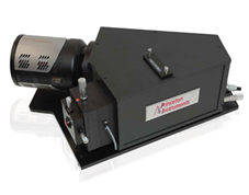
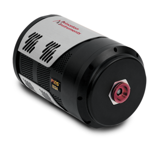
In their measurements the researchers use a setup scanning a fiber optic probe over tissue samples. The probe design contains fibers for FLIM and Raman excitation and signal collection as well as filters required in Raman spectroscopy for clean laser excitation and suppression of elastic scattered light. As FLIM is the faster of the two techniques, it is used to scan the surface and identify targets (detecting changes in fluorescent lifetime) to be investigated using Raman spectroscopy by excitation with a 785nm multimode laser. The Raman signals are obtained using a LS-785 spectrograph and PIXIS camera.
Advanced numerical techniques (extended multiplicative signal correction EMSC, partial least-squares linear discriminant analysis PLS-LDA) are used for systematic background subtraction and data analysis. The changes in the Raman spectra correctly identify cross-linked from non-cross-linked tissue and provide the researchers high biochemical sensitivity to identify calcification, while FLIM is shown to better detect the degree of cross linking in the collagen matrix.
The authors conclude that “FLIM and Raman imaging has a strong potential to address some of the pressing needs for nondestructive and label-free monitoring of tissue engineering processes and their applications in regenerative medicine.” Combining both measurement technique leads to a higher understanding of the biochemical and structural processes of BP, helping in studying the performance of the tissue over time.