Angela Belcher
Scientific Reports
Deep-tissue optical imaging of near cellular-sized features
Introduction
Researchers from MIT, in collaboration with Koch Institute for Integrative Cancer Research, published a breakthrough scientific report in biomedical engineering. They designed & developed an NIR optical imaging system named DOLPHIN, which stands for “Detection of Optically Luminescent Probes using Hyperspectral and diffuse Imaging in Near-infrared.” Their system holds promise for high-resolution, deep-tissue imaging. DOLPHIN achieves the following:
- • Resolution of probes through up to 8 cm of tissue phantom
- • Identification of spectral and scattering signatures of tissues without a priori knowledge of background or autofluorescence;
- • 3D reconstruction of live whole animals.
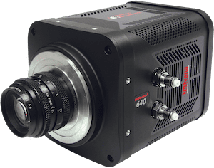
For hyperspectral imaging (HSI) mode, they utilize a liquid nitrogen cooled InGaAs camera (OMA:V 2D, 256 × 320 pixel array used without binning, detection range: 900-1,700 nm, temperature –100 °C) at the exit slit of monochromator equipped with a 150 g/mm NIR diffraction gratings. To the best of their knowledge, DOLPHIN is the first demonstration of utilizing both HSI and HDI modes in a trans-illumination configuration to investigate NIR-II fluorescent signals. In contrast, previous HSI and HDI technologies worked mainly in the visible and NIR-I wavelengths, utilized either epiillumination or reflectance configurations that result in shallower depths of detection, and also relied on mapping to reference spectra to identify features of interest.