Displays made of Organic Light Emitting Devices (OLEDs) have found widespread use over the past few years in mobile, TV and computer screens. Compared to other technologies OLEDs emit visible light directly so no back lighting is necessary, leading to darker black levels and improved contrast ratios while being thinner and more easily manufactured.
Current research aims to find new and improved OLED materials with improved efficiencies and device lifetimes. In OLEDs, the light emitting material is embedded in a host material. This plays an important role in maximizing luminescent efficiency as well as charge and exciton transport. However, the host material building the emissive layer in OLED devices often plays a significant role in the stability and lifetime and seemingly small chemical changes can have large effects.
Researchers from University of Minnesota and DuPont Electronics, from the Russell Holmes laboratory, recently investigated the molecular mechanisms influencing lifetime of OLEDs using mass spectrometry and a range of optical and spectroscopic techniques. The study uses two host materials, CBP and CDBP of similar molecular structure, however lifetime in CDBP devices is decreased by a factor of 10 or more due to formation of excimer states.
Optical and spectroscopic techniques provide a lot of useful information to investigate this observation, and spectral information was obtained using a IsoPlane 81 (previously knwon as the FERGIE) spectroscopy system. The lifetime of the devices can be monitored by quantitative measurement of the low temperature photoluminescence and electroluminescence signal over several hours. These measurements are performed with low laser excitation powers to avoid laser induced degradation effects and require sensitive detection for the PL signal.
The spectral shape of the thin film phosphorescence gives information about the material composition as well as the formation of the excited states by looking at the spectral signature of the excimer states and excimer/monomer signal ratio.
High fidelity EUV and X-ray radiation is important in many applications such as medical and material X-ray radiography, X-ray diffraction. Most commonly the high energy radiation is generated by accelerating charges in magnetic fields (for example in synchrotron facilities).
Another approach is using inverse Compton scattering where the magnetic field is replaced by an electromagnetic wave from a laser pulse. This process requires less effort to produce high energy radiation, but current sources have to be optimized further to produce higher quality output beams.
Researchers from Germany present an approach where they measure the spectral shape of the X-ray beam to characterize the radiation source. They use an in vacuum CCD camera to record the spectra by counting single photon events.
An international team of researchers centered in Germany designed and built a new EUV microscope with sufficient magnification for imaging of scattering and absorption of plasmas created with sub picosecond EUV pulses created from cryogenic hydrogen jets. The EUV radiation is created in a free electron laser (FEL, FLASH in Hamburg Germany) built to provide short pulsed, high quality radiation at extremely short wavelengths.
Understanding the physics of plasmas has important applications in other fields. Higher resolution of plasma dynamics will improve understanding and modelling of processes in planetary science and fusion research. EUV light can penetrate deeply into plasmas (unlike visible light) so it is well suited to study inner workings of plasmas.
For their setup the team created proper objective, designed for right magnification (practical FOV size with highest resolution given 13.5 μm pixel pitch of CCD). A sensitive CCD detector allows for fast imaging of scattering from single radiation pulses.
Particle accelerators and synchrotrons are our most perfect and brilliant light sources of visible to x-ray radiation for scientific research and development, from material to life science. Recently techniques using strong laser fields in plasmas have emerged as an alternative for acceleration because of their much smaller size (<1 meter vs. 100s of meters for synchrotrons).
However, the emerging particle beams still have significant quality issues compared to the currently used beams. Researchers at the Synchrotron SOLEIL in France report on work of improving the beam quality where they monitor the quality and shape of the radiation that is produced from them. Vacuum compatible scientific CCDs allow for sensitive detection of radiation from UV to soft x-rays in this application.
Researchers around Jens Biegertin, in Spain are investigating real time behavior of phase transitions using X-Ray spectroscopic techniques.
X-Ray spectroscopy can give element specific information about electrons in a material as well as determine information about the structure (how far are atoms apart). The researchers apply new light sources using high harmonic generation that produce ultrashort attosecond length pulses that span a wide energy range over hundreds of eV in the soft X-ray regime (specifically in the water window a region from around 280eV to 530eV where water is transparent, but carbon and organic materials are not).
They are able to correlate the electronic information and material structure in real time and want to apply this knowledge for better understanding of phase transitions for solids, liquids and gases.
Diffraction gratings are used as the dispersive element in spectral analysis from the IR to the soft X-Ray range. A common problem in any wavelength range is the influence of higher diffraction orders that can either overlap with the main signal or complicate calibration of the measurement instruments. A research team in China is developing new grating types based on nanotechnology fabrication techniques that show much larger suppression of higher diffraction orders than blazed diffraction gratings.
Interestingly the new gratings do not have a periodic, repeatable structure, but are made of millions of nanometer sized pillars arranged in a quasi-random fashion. To test the gratings in an X-Ray monochromator, the researchers used a sensitive CCD (PIXIS-XO) that detects very low signals by integrating for extended amounts of time. While the suppression of higher orders is very high in the nanopillar gratings the overall efficiency is low (however it is still comparable to the efficiency achieved by conventional methods of higher order suppression).
This featured article highlights how the FERGIE (now IsoPlane 81) system is being utilized in material science research. Researchers from Birck Nanotechnology Center, Perdue University report on the electrocaloric effect (ECE) of a new electrocaloric (EC) material CIPS, which is CuInP2S6.
EC materials are potentially useful for EC refrigerators. These EC refrigerators have low noise and are environmentally friendly. They are also able to be scaled down to small dimensions leading to nanorefrigerators. Thus, ECE is promising for future cooling applications, especially in the micro- to nanoscale such as on-chip cooling.
The researchers characterized their novel material via temperature dependent Raman spectroscopy, utilizing a 100x 0.75NA objective integrated to FERGIE system. A single mode fiber coupled 532 nm laser with a <1 MHz bandwidth was used as the excitation source.
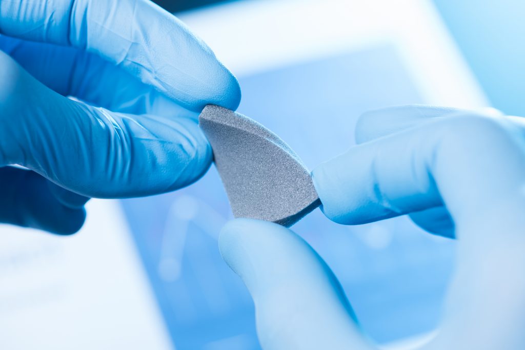
In an environment of constantly changing health challenges, analytical techniques play an important role in the development of new and enhanced drugs as well as the quality control of drug production. Among a wide range of techniques, Raman spectroscopy has an important place for material analysis. It is a non-invasive and non-destructive technique that only requires small efforts of sample preparation. Materials and molecules can be identified with high specificity by their Raman fingerprint. Raman spectroscopy is a good tool for investigating the chemical and structural properties of materials as well.
Researchers at the New Jersey Institute of Technology use Raman spectroscopy for experiments on particle materials and nanotechnology for improved drug delivery. One challenge the in the development of new drugs is their poor water solubility. The human body is made of up to 60% of water and high drug solubility is often an important factor for effective a drug can be applied.
In a recent publication the researchers report on experiments to enhance the solubility of an anti-fungal drug call Griseofulvin. They prepare dispersions of the drug using nanocrystals, polymers and surfactants and investigate their structure and composition. Raman spectra were taken using a FERGIE system at 785nm laser excitation. The position of Raman bands and their width gives insights into the crystalline or amorphous structure of the drug as well as its interactions with the other materials. In combination with calorimetric and X-Ray diffraction techniques Raman spectroscopy helps the researchers to better understand how well their samples mix with water.
Raman spectroscopy is a highly sensitive tool for material identification and identifying structure of materials down to molecular levels. It is a ubiquitous tool not only in fundamental research but applied research as well. Mark Waterland from Massey University and his team are applying Raman spectroscopy to solve complex analytical problems across research fields such as medicine, ecology, food science and more.
Here are a few examples studied by the group over the past few years:
• Improving the quality of road pavements and understanding their failure mechanisms. The bitumen/aggregate mixtures used for road pavements are poorly compatible due to the hydrophilic nature of the aggregates. Raman spectroscopy allows for the analysis of the molecular structure of the surfaces of these materials down to the existence of specific bonds such as O-H or P=O as these emit Raman signals at different wavelength bands. This can lead to a better understanding of binding interactions and how to improve them.
• Quality control of leather. There is a significant amount of loss in leather manufacturing due to a quality defect called looseness. The defects are typically only identified after the raw material has undergone a series of resource intensive processing steps. Raman spectroscopy can help identify materials prone to showing defects early in the process. Once the spectral features for identification are known they can be combined with a chemometric model for analysis and classification.
• Finding contamination in milk. Surface enhanced Raman scattering can identify specific contamination molecules, potentially even single molecules. However, it is hard to apply to a colloidal liquid such as milk due to the strong light scattering and nature of SERS samples when applying standard sample preparation which make SERS signals hard to detect. Using a new sample preparation process, the researchers are able to achieve detection of very low concentration of contaminants.
The work of Mark Waterlands team clearly shows the high impact of Raman spectroscopy for applied research problems.
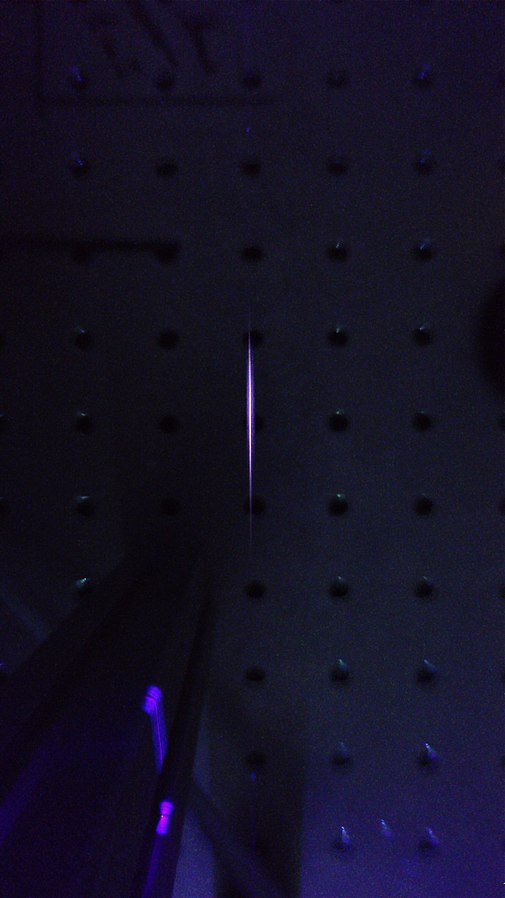
High power lasers can show non-linear optical effects that lead to the self-focusing of laser beams, producing thin channels of plasmas along the laser propagation path which are referred to as laser filaments. Scientists believe that understanding the dynamics and processes in these filaments could build the basis for applications in power transmission, as plasma antennas or long-range sensing.
courtesy: Jean-Christophe, Michel, Delagnes
Researchers around Ruxin Li from the State Key Laboratory of High Field Laser Physics are investigating the interaction of laser filaments with strong electric fields and optical spectroscopy. This plays an important role by measuring the fluorescence of Nitrogen in the laser plasmas. A pair of imaging lenses are used to guide the emitted radiation into the entrance slit of a SpectraPro HRS spectrograph, providing high resolution to observe the spectral features of the plasma ions.