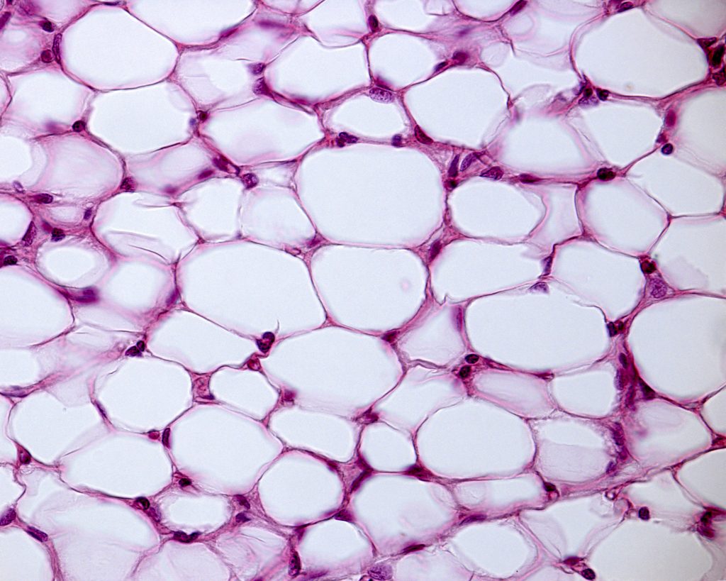
NIR-II in vivo and in vitro imaging enable detailed observation of blood vessel growth in enhanced adipose tissue scaffolds.
Defects in soft tissue, often caused by burns, congenital deformities, and tumor resections, have long been a common difficulty within reconstructive surgery. Engineered, vascularized adipose tissue has been extensively studied as a potential aid for repair of soft tissue damage or defects. Typically, when utilizing adipose tissue scaffolds, adipose-derive stem cells are introduced to promote tissue regeneration and healing of the soft tissue defect.
However, adipose tissue scaffolds are limited as typically they display delayed neovascularization, the process in which new blood vessels are formed, and unstable adipose formation. Researchers from China have begun tackling these limitations by introducing CCL2, an exogenous chemokine,which enhances both adipose tissue and blood vessel growth. To counteract the effects of delayed vascularization, the researchers also introduced a vascularized fibrous capsule to promote neovascularization.
The researchers studied the effects of CCL2 alone and CCL2 and the capsule in both in vitro and in vivo environments. In order to visualise neovascuarlization in vivo, the researchers obtained NIR-II fluorescence images with a NIRvana camera. They used PbS QDs which were administered immediately before images were acquired. These QDs are advantageous as they have very good biocompatibility and fluoresce well within the NIR-II window, at approximately 1300 nm. This wavelength is beneficial for in vivo imaging as it allows for deeper penetration depth with less scattering and lower tissue autofluorescence, resulting in better image quality.
However, these wavelengths are unable to be imaged using traditional silicon cameras, such as CCD or sCMOS. Instead InGaAs cameras, such as the NIRvana camera, must be used. In addition, the NIRvana InGaAs camera has deep cooling and ultra-low dark current, optimized for long acquisition times typical in in vivo imaging. Furthermore, the high quantum efficiency of the NIRvana means it has high infrared sensitivity for faint signals common when imaging in vivo blood vessels. The researchers found that the use of CCL2 and the capsule provided the highest percentage of blood vessels, with strong indication that new blood vessels had developed.
Fluorescence imaging helps to characterize hydrogels manufactured for in human tissue.
There is a growing need for micro- or nanostructures machines that mimic dynamic human tissues for applications such as drug delivery, tissue engineering, and nanomechanics. Although this area of research has previously been focused on 3D-printed constructs, they are limited by their bulky size and rigidity. Hydrogels, however, are an emerging alternative and have shown promise in many applications such as cancer treatment and wound healing as they act similarly to human tissue. In addition, hydrogels have good biocompatibility and degradability, with the ability for cell adhesion and growth.
To fully utilize hydrogels for these applications, controllable stimuli-responsive behavior is essential. Additive manufacturing methods have been shown to produce stimuli-responsive behavior in a controlled way. In addition, additive manufacturing allows for a 4D construct, which takes advantage of time-dependent behavior of stimulus-responsive materials such as hydrogels.
Researchers at Jiangsu University, Huazhong University of Science and Technology, and Johns Hopkins University used two-photon polymerization, a typical additive manufacturing method, with a femtosecond laser to create hierarchical microstructures and nanostructures. These structures created a controllable surface tension mismatch. They were able to show that hydrogels with this controlled stimuli-responsive behavior were able to focus a bright spot of light by swelling and shrinking, morph in shape in a similar manner to how plants move, and deform by absorbing localized light energy.
Fluorescence imaging was one method of nanostructure characterization used by the researchers. Fluorescence images were used to determine the structures of the fabricated hydrogels. The IsoPlane and KURO were also used to image the biocompatibility of the hydrogels. To test this, active cells were introduced to the hydrogels, and fluorescence images indicated that the active cells crawled onto the prepared surface, indicating desirable adhesion and biocompatibility for cells.
The reflected fluorescence images of the hydrogel structure were captured using an IsoPlane-320 spectrometer and KURO sCMOS camera. The IsoPlane-320 is ideally suited for sensitive micro- and imaging spectroscopy measurements. The advanced optical design of this spectrometer make it possible to achieve higher spatial localization of the measurement signal and reliable measure spectra of several points simultaneously with clear separation of spectral signals. The IsoPlane is also able to capture broadband as well as high resolution spectra with a lot of spectral details. The KURO sCMOS camera features a scientific, back-illuminated CMOS sensors. The high quantum efficiency of the sensor enables measurement of weaker signals with higher S/N ratio. The low readnoise of the camera is ideal for applications where many spectral images have to be read out with high measurement rates.
Cardiovascular disease is one of the most common causes of death worldwide. One of the typical indicators of this disease is calcifications within the coronary artery. The most established method for measuring these calcifications is computed tomography (CT). Within the clinic, these CT scanners rely on energy-integrating detectors, which utilize a scintillator to convert high energy photons into visible light. However, they are limited in spatial resolution which can result in either an overestimated diagnosis or an underestimated one. In comparison, photon-counting detectors, currently not used in clinic, use a semiconductor to convert x-ray photons to electrical signals, allowing for smaller signals and improved spatial resolution.
Researchers from Linköping University and the Mayo Clinic in Rochester (USA) have compared a photon-counting detector CT with an energy-integrating detector CT to determine which method is more accurate at quantifying calcifications on the coronary artery. They relied on micro-CT measurements as the reference standard.
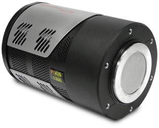
These researchers found that the photon-counting detector CT was not only able to quantify coronary calcifications much more accurately, but also produce lower noise than the standard energy-integrating detectors. The researchers utilized a PIXIS-XB within their micro-CT set up, creating a custom-built micro-CT scanner to create the standard reference. By accumulating hundreds of images over 360° sample rotation they were able to reconstruct the sample for referencing.
In order to calibrate x-ray detectors, they need to be tested at how well they respond to different x-ray energy levels at dedicated x-ray test facilities. Researchers from China developed a hard x-ray test facility to test various hard x-ray detectors based on a single crystal monochromator.
They utilized 3 single crystals within the monochromator to cover the energy range 21-301 keV. The system was comprised of 4 parts, an x-ray source, a collimating structure, a single crystal monochromator, and two detectors. These detectors were a high-purity Germanium detector, and the PIXIS-XF CCD camera.
The researchers found there to be a good linear relationship between the x-ray tube current and the monochromatic light flux. However, the energy resolution worsened with increasing energy value, as the spectra broadened. Overall, they found the x-ray test facility to accommodate a wide energy rage with good stability, and therefore suitable for multiple testing experiments.
The low noise of the PIXIS-XF made it a great option for monitoring the x-ray light spot. The researchers found the dual speed operation suitable for both steady state as well as for high speed applications, and therefore an ideal option for monitoring their new system.
X-ray mapping is an important tool for non-destructively assessing the chemical composition of solid samples. With the recent development in both computational technology and detector technology, hyperspectral x-ray maps can now be used to further analysis.
Soft x-ray emission spectrometers are one of the most recent hyperspectral detectors, and can measure very low energy x-rays, increasing chemical analysis through the use of direct lithium measurements and spectral mapping of L, M, and N lines of certain elements.
Typically, these spectrometers utilize a 2048 x 2048, x-ray sensitive CCD camera for measurement. However, CCDs of this size are limited by the amount of time taken to readout a spectrum, even with on-chip binning.
Researchers from Australia attached a SOPHIA-XO onto a soft x-ray emission spectrometer, alongside other detector types, to optimize the experimental configuration of x-ray hyperspectral mapping. With the four-port readout of the SOPHIA-XO, the researchers were able to achieve readout speeds 10x faster than that of traditional CCD cameras. The researchers were concerned that the faster readout would result in a lower signal-to-noise ratio, due to the reduction of signal accumulation. However, the improved electronics of the SOPHIA-XO improved the noise characteristics, increasing the signal-to-noise ratio in comparison to other models.
X-ray free electron lasers (XFELs) are a great source of coherent, intense radiation achieving sub-angstrom wavelengths. They are advantages in a range of applications from chemistry to structural biology. However, they are costly, large and require advanced technology to achieve these radiation beams.
Laser wakefield accelerators (LWFAs), which accelerate particles to high energies by utilizing the enormous electrostatic field of an excited plasma wakefield, are able to produce radiation over just a few millimeters – centimeters. This makes them a promising compact, economically viable alternative to XFELs for laboratory settings.
However, the relatively poor quality of electron beams based on LWFAs makes them challenging, as typical FEL configurations rely on a high-quality, stable electron beam. Researchers from the Chinese Academy of Sciences have demonstrated an FEL using a LWFA as proof-of-principle. Through the use of an LWFA accelerated electron beam, they are able to generate undulator radiation with an exponential amplification. This radiation is centered on 27 nm, with a maximum photon number of ~1010 per shot.
In order to evaluate the on-axis undulator radiation produced, the researchers used a SOPHIA-XO. By measuring the number of photons collected by the SOPHIA-XO, they were able to determine the energy of the undulator radiation. The researchers also installed a transmission grating in front of the SOPHIA-XO to spectral diagnostics of the radiation.
Ultra-High-Speed, Time-Resolved SRS Spectroscopy in CombustionApplication Notes
Novel (3D) Neutron Imaging Technique for Nondestructive Testing Made Possible by Large-Area, Intensified CCD Camera System
Introduction
In combustion, until recently only two temporal optical gating schemes were available to increase signal-to-noise ratio (SNR) for time-resolved spontaneous Raman scattering (SRS) spectroscopy. Problematic optical background noise could be rejected either by electronic gating with an image intensifier or by using a mechanical shutter. Unfortunately, each of these traditional approaches has its shortcomings.
Image intensifiers, for example, provide excellent optical background noise rejection via <2 nsec gating capability but carry several inherent limitations, such as lesser image quality and lower dynamic range. On the other hand, while a high-speed mechanical shutter with a rotary optical chopper is able to deliver wider dynamic range without diminishing the quantum efficiency (QE) of the detection system’s CCD, its 30 Hz speed and ~10 µsec gating are not sufficient for rejecting noise and can result in transmission losses of up to 50%.
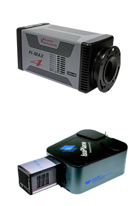
In 2010, Dr. Jun Kojima of the Ohio Aerospace Institute, working with Dr. David Fischer and Dr. Quang-Viet Nguyen (NASA Glenn Research Center), described an architecture* for SRS that employs a frame-transfer CCD operating in a subframe burst-gating mode to realize time-resolved combustion diagnostics.1 This patented technique enables all-electronic optical gating at microsecond shutter speeds (<5 µsec) without compromising optical throughput or image fidelity. Dr. Kojima uses a Princeton Instruments ProEM® electron-multiplying CCD (EMCCD) camera for this method.
When utilized in conjunction with a pair of orthogonally polarized excitation lasers, the technique described above measures single-shot vibrational Raman scattering that is minimally contaminated by optical background noise. Nonetheless, its relatively long gating (~5 µsec) still leaves room for improvement in terms of optical background rejection.
Recently, Dr. Kojima has developed another advanced technique for measuring time-resolved SRS spectroscopy in combustion (see Figure 1). An overview of this new approach, which offers evenhigher SNR and permits ultra-high-speed observation of combustion dynamics, is provided herein.
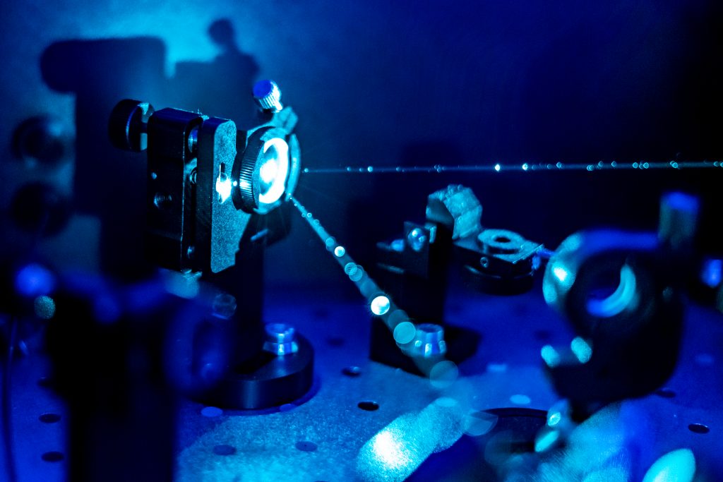
Figure 1. Dr. Jun Kojima of the Ohio Aerospace Institute testing new technique at NASA Glenn Research Center.
New Way To Measure Time-Resolved Spontaneous Raman Scattering
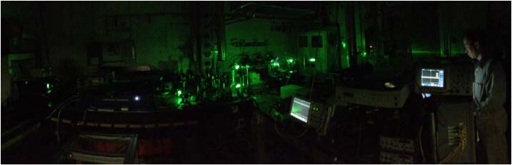
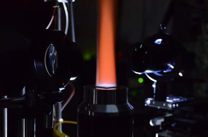
Dr. Kojima’s new experimental apparatus is shown in Figure 2. It measures Raman scattering with much faster gating (<2 nsec), wider dynamic range, and higher sensitivity in combustion than previously reported techniques, allowing observation of flame instability dynamics via a newly introduced Princeton Instruments intensified emICCD camera that keeps up with the latest 10 kHz lasers.
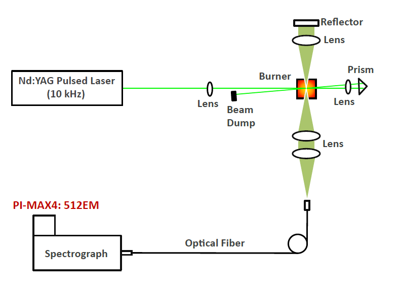
Figure 2. Experimental apparatus showing a high-speed laser Raman diagnostic system employing an Nd:YAG pulsed laser (532 nm, 88 nsec pulse width, 10 kHz repetition rate) and a Princeton Instruments PI-MAX4:512EM camera coupled to a lens spectrograph. Courtesy of J. Kojima (OAI/NASA).
Here, a second-harmonic Nd:YAG pulsed laser was used as an excitation source (200 W) and operated at a 10 kHz repetition rate to interrogate a flame. The scattering light was collected by fiber-coupled lens optics and transmitted to a volume-transmissive lens spectrograph equipped with a PI-MAX®4:512EM camera from Princeton Instruments.
To enhance the SNR, the image intensifier was operated at a 10 kHz rate to keep pace with the high-speed laser and gated at 90 nsec to cut out the optical flame emission background, while the EMCCD was operated at a rate of 1 kHz (i.e., 10 laser-shot accumulation) using a special feature within Princeton Instruments LightField® software that enables customization of CCD size and readout speed. This custom detection setting effectively enabled the diagnostic system to achieve the highest signal level ever achieved in the NASA facility without sacrificing the necessary kHz data rate.
Enabling Technology
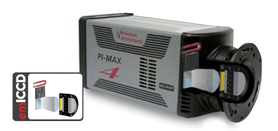
New PI-MAX4 emICCD (see Figure 3) cameras from Princeton Instruments leverage the key advantages of both EMCCDs and ICCDs by fiber optically coupling an EMCCD to an image intensifier. This innovative emICCD technology lets the new cameras deliver an unrivaled combination of precision, true single-photon detection, intelligence, and speed.
Figure 3. The new Princeton Instruments PI-MAX4:512EM is the first scientific camera on the market to utilize revolutionary emICCD technology. The PI-MAX4:512EM uses a front-illuminated EMCCD; the PI-MAX4:512EMB uses a back-illuminated EMCCD.
The PI-MAX4 emICCD camera’s back-illuminated EMCCD boasts 95% peak QE for the highest signal throughput of any ICCD camera. Furthermore, coupling the EMCCD to an image intensifier via fiberoptics delivers 6x higher light throughput between the image intensifier and the detector than lens-coupled configurations. As a result, emICCDs provide the highest signal-to-noise (SNR) of any gated imaging and spectroscopy detectors.
The exceptional linearity and dynamic range of these new emICCD cameras, achieved by intelligently programming gains between the image intensifier and the EMCCD, are critical for quantitative imaging and spectroscopy applications such as combustion. Their true single-photon detection capability, meanwhile, ensures the high sensitivity needed for light-starved applications. An oscilloscope-like user interface (see Figure 4) even remembers complete experimental setups. In addition, thanks to the ability to acquire 10,000 spectra/sec when operated in a special custom chip mode, Princeton Instruments’ emICCD cameras can capture every pulse from next-generation lasers.
Figure 4. An oscilloscope-like user interface enhances the utility of Princeton Instruments’ emICCD cameras.
Results
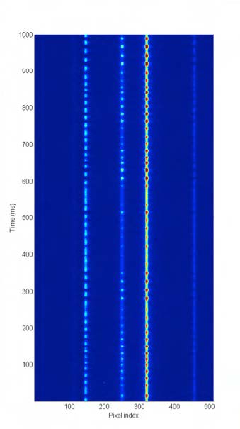
High-speed laser spectroscopy measurements were performed using the diagnostic apparatus shown in Figure 2 at the Atmospheric Pressure Combustion Diagnostics (APCD) lab, NASA Glenn Research Center, in Cleveland, Ohio. Figure 5a shows a close-up of a flame. Figure 5b presents data for temporal variation of spontaneous Raman scattering of combustion species (oxygen, nitrogen, and water vapor) measured at the tip of a fuel-lean hydrogen-air flame. Pixel region (1 to 512) corresponds to the wavelength region of 486 to 680 nm.
Figure 5a. Close-up of flame.Figure 5b. Time-series Stokes Raman scattering spectra recorded at a sampling rate of 1 kHz over one second in an oscillating fuel-lean hydrogen-air flame using the high-speed Raman diagnostic apparatus with PI-MAX4:512EM. Courtesy of J. Kojima and D. Fischer (OAI/ NASA).
The signal visibility in Figure 5b is significantly higher than previously reported data of this kind. It is clearly seen from the data that the flame oscillates at a certain frequency (here, ~46 Hz) due to an interaction of the flame with ambient air entrainment.
A careful observation reveals that the pure rotational band is in inverse correlation with the O2 and N2 spectra. This is explained by the fact that the pure rotational band is a flame marker (high temperature) in contrast to the two species, which decrease their peak intensity at higher temperature. Higher concentration of water vapor (H2O), the combustion product, appears when the flame is observed. It is significant that flame dynamics were characterized in a species-resolved fashion using high-speed Raman spectroscopy.
Future Directions
The recent use of a new diagnostic apparatus to measure the dynamics of each individual molecular species, as opposed to simply acquiring bulk information (e.g., pressure), points to the possibility of performing temperature and frequency analyses of species in combustion. Ultimately, such potential applications could become diagnostic tools for fuel-air ratio dynamics at different temperatures and pressures.
Advances in scientific detector technology, such as the PI-MAX4 emICCD camera’s ability to gate out all optical background noise at <1 nsec and thus improve SNR for time-resolved spontaneous Raman scattering spectroscopy in combustion, continue to extend the horizons of research in this area.
Reference
J. Kojima, D. Fisc her, and Q .-V. Nguyen, Opt. Lett.
35, 9 (2010).
Measuring Fusion Plasmas Using SpectroscopyCustomer Stories
Ted Biewer
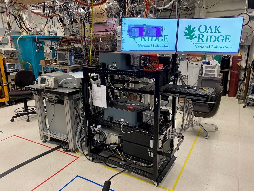
Oak Ridge National Lab, Fusion Energy Division, Diagnostics and Control
Background
The fusion diagnostics and control group led by Ted Biewer at Oak Ridge National Laboratory specializes in measuring and monitoring properties of plasmas in fusion experiments. We talked to Drew Elliott, a scientist in the group: “What we do in our group is develop and operate diagnostics to better characterize a lot of these experiments.” While several groups and experiments worldwide are searching and developing ways to create more efficient and economical fusion experiments, measuring key parameters of fusion plasmas remains challenging. “We are diagnostic experts and are able to make more accurate measurements or make new measurements which other groups either don’t have the personnel, hardware or expertise.”
In an effort to bring their expertise to multiple fusion labs around the world, the researchers recently developed a compact and portable system that can be transported and operated at different experiment sites. The instrument implements a technique called Thomson scattering (scattering of electromagnetic radiation by charged particles) which Dr. Elliott explains as basically being a very accurate way to measure temperature or density in hot plasmas. Using optical methods is crucial in this process as physical probes would be destroyed in the hot and harsh environments found in fusion plasmas.
Using their new setup the group will be able to support experiments worldwide by giving them diagnostic capabilities and information that they did not have before.
Challenge
The main challenge in developing the new setup was that the spectroscopy system needed to be compact enough to be transportable, but also have flexibility to adapt to a wide range of experiments that could have very different parameters. For example, plasma temperatures might range from relatively cold plasmas (at around 1000 times room temperature) to temperatures approaching those inside of the sun. Dr. Elliott explained that the temperature limit that can be measured using their spectroscopic technique essentially depends on the spectral bandwidth of the spectrograph which should be able to adapt its spectral range for different experimental situations.
One problem, encountered in conventional spectrographs is that their instrument response function changes significantly between the center and edges of the camera sensor due to optical aberrations. Spectral lines would then change size and shape depending where on the sensor they are observed. The team required a better instrument response so complex and difficult corrections in data processing could be avoided.
Experiments on plasmas also require detectors capable of very short exposure times to suppress the very bright background radiation of the hot plasma. If the exposure is timed precisely with the signal from a short laser pulse, this background light can be effectively negligible compared to the laser signal.
“Overall, the IsoPlane and PI-MAX combination allowed us to set up a compact system that achieves the required measurement capability”
Solution
The group implemented a spectroscopy system consisting of IsoPlane-320 spectrographs and PI-MAX4 ICCD cameras for their transportable diagnostic system. The IsoPlane-320 has an advanced optical system, compared to conventional lab spectrographs, that minimizes optical aberrations. As a result, the instrument response and resolution remain constant across the detector. Low aberrations also allow for observation of a larger number of spectral tracks. Thomson scattering experiments often use multiple fiber optic inputs, addressing different locations in the experiment. This spectrometer and camera configuration allows the Oak Ridge team to observe all these channels simultaneously. IsoPlane spectrographs also come equipped with a triple grating turret, so the spectral bandwidth can be changed quickly by switching to gratings with larger or smaller groove density.
Moreover, the PI-MAX4 intensified CCD camera allows for nano-second gate times, necessary to suppress the bright plasma background. The gate opening can be synchronized with picosecond precision to the signal induced by the fast laser pulses.
“Overall, the IsoPlane and PI-MAX combination allowed us to set up a compact system that achieves the required measurement capability.”
High-Harmonic Generation (HHG) and Highly-Sensitive Scientific Cameras for Soft X-ray ApplicationsApplication Notes
Introduction
As is the case with many scientific and commercial technologies, the x-ray imaging and spectroscopy instruments utilized to perform leading-edge academic and industrial research are becoming smaller, more cost effective, and — in a sense — more personalized. For years, researchers across a wide range of disciplines whose daily work relies upon soft x-ray energies have had to schedule sessions at large, high-brilliance x-ray sources in order to conduct critical experiments.1 Time at these expensive-to-operate shared resources (e.g., third- and fourth-generation synchrotrons and x-ray free-electron lasers) remains a precious commodity.
The advent of high-harmonic generation (HHG) instruments, however, is beginning to reshape the landscape for researchers desirous of performing various imaging and spectroscopy experiments in the soft x-ray energy regime. HHG light sources complement large-scale synchrotrons and x-ray free-electron laser (XFEL) sources; among the key advantages of using tabletop HHG light sources for x-ray applications are easier access, full synchonizability to the sub-femtosecond level, and attosecond temporal resolution.2,3
Advanced electronics, nanoscience, semiconductor, materials science, polymer science, biotech, and life science applications can benefit from pairing tabletop HHG light sources with x-ray imaging/spectroscopy detectors to facilitate experimental techniques such as coherent x-ray diffraction imaging (i.e., lensless x-ray imaging), x-ray absorption fine-structure (XAFS) spectroscopy, near-edge x-ray absorption fine-structure (NEXAFS) spectroscopy, and extreme ultraviolet (EUV) spectroscopy.2–7
This application note will address recent work performed by researchers in the Attoscience and Ultrafast Optics group of Prof. Dr. Jens Biegert at the Institute of Photonic Sciences (ICFO) who developed/utilized a tabletop attosecond HHG light source in concert with a Princeton Instruments x-ray CCD camera to demonstrate NEXAFS spectroscopy at the socalled water window’s carbon K-edge of a polyimide foil and retrieve the specific absorption features corresponding to the binding orbitals of the carbon atoms in the foil.4,5
Experimental Setup
The K-absorption edges of carbon, nitrogen, and oxygen occur within what is often referred to as the water window, a range of wavelengths (2.3 nm – 4.5 nm) at which water is virtually transparent. Because each of these elements is fundamental to life, the ability to make increasingly more sophisticated observations in the water window is critical to leading-edge research being conducted by biologists, chemists, and physicists across many diverse fields.5
Until recently, such observations relied on the utilization of large-scale, high-brilliance sources. The aforementioned research group from ICFO in Barcelona is among a number of groups around the world2,6 who are implementing tabletop HHG instrumentation as a compact, cost-effective alternative.
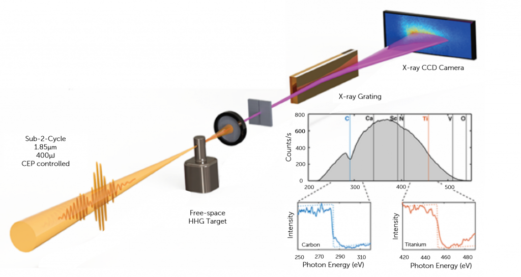
The ICFO group, for example, has developed a tabletop HHG light source to perform high-resolution x-ray absorption spectroscopy on a polyimide foil. Their source rests on a highly stable kilohertz laser system that drives an optical parametric amplifier with subsequent hollow-core-fiber pulse compression, and results in sub-2-cycle carrier-envelope-phase–stable (CEP-stable) laser pulses with a central wavelength of 1.85 μm; the high harmonics generated by this source have reached photon energies up to 535 eV, far beyond the carbon K-edge.4,5
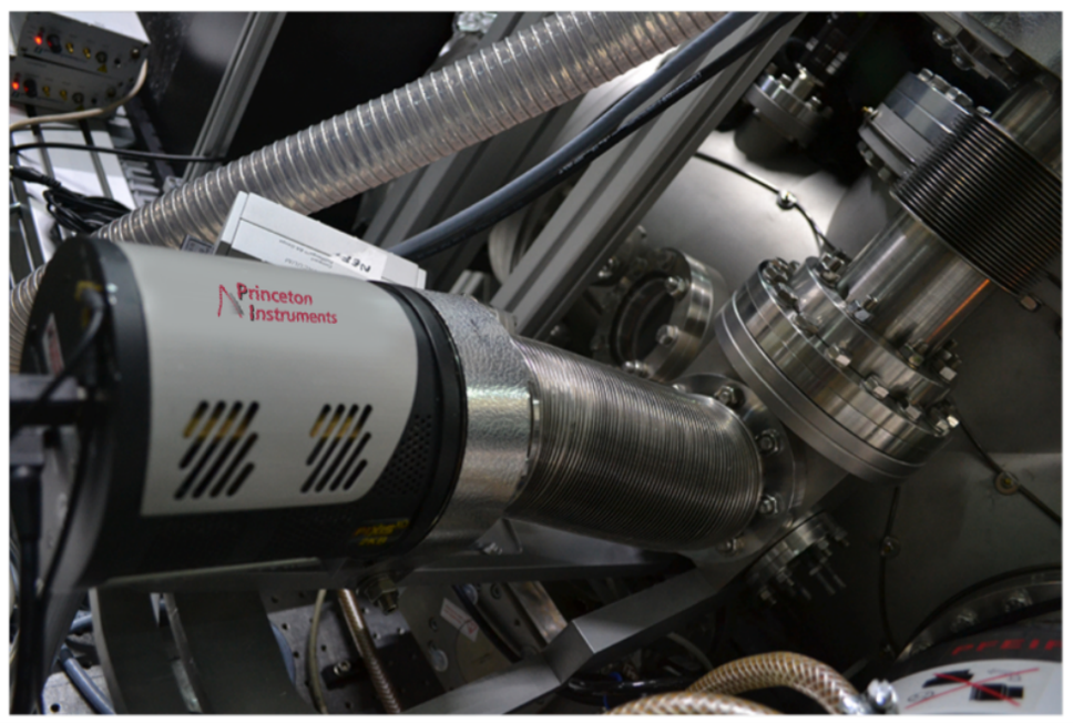
The group used second-harmonic-generation frequency-resolved optical gating (FROG) to measure compressed pulses, and achieved bright soft x-ray flux and a 535 eV cutoff in the water window by focusing the CEP-stable sub-2-cycle pulses with an f = 100 mm silver-coated curved mirror into the HHG target. The harmonic spectra were resolved with an x-ray spectrograph and a cooled Princeton Instruments PIXIS-XO x-ray CCD camera.4 Refer to Figures 1 and 2. The researchers measured the resolution of their x-ray spectrometer as 0.25 eV at 300 eV.
Figure 1: Illustration of the ICFO research group’s experimental setup for NEXAFS spectroscopy using a tabletop HHG light source and a scientific x-ray CCD camera. Courtesy of Prof. Dr. Jens Biegert (Institute of Photonic Sciences, Barcelona).
Figure 2: Photo of the experimental setup illustrated in Figure 1 incorporating a Princeton Instruments PIXIS-XO camera. Courtesy of Prof. Dr. Jens Biegers (Institute of Photonic Sciences, Barcelona)
Results
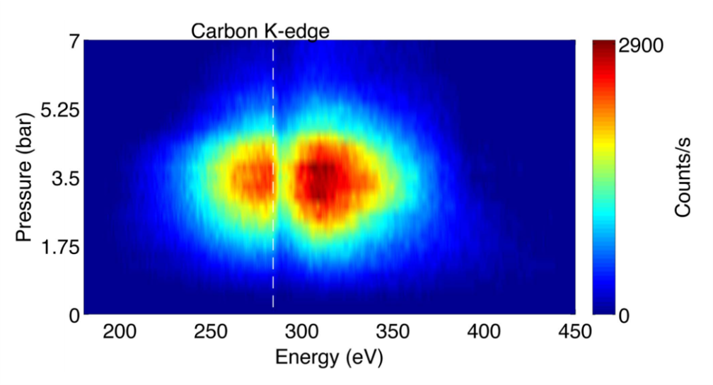
The researchers at ICFO optimized harmonic flux by scanning the target-backing pressure from 0 to 7 bar of neon. Figure 3 shows the results of integrating the HHG spectrum (for a given backing pressure) for 5 sec in steps of 0.25 bar per spectrum; it clearly illustrates that a cutoff far beyond the 284 eV carbon K-edge was achieved and that the highest yield was reached (together with the highest cutoff) at a backing pressure of 3.5 bar.4
Figure 3: HHG spectra as a function of neon backing pressure. The highest harmonic yield coincided with the highest cutoff at a backing pressure of 3.5 bar (4). A PIXIS-XO camera was used to acquire the data. Courtesy of Prof. Dr. Jens Biegert (Institute of Photonic Sciences, Barcelona). First published in Opt. Lett. 39, 5383-5386 (2014).
After determining the optimum pressure for the target, CEP control of the spectral shape was investigated by measuring x-ray beam brilliance and photon flux. This control was demonstrated to be crucial for avoiding potential over-averaging of pre- and post-edge structures that vary from shot to shot while integrating a NEXAFS spectrum.4
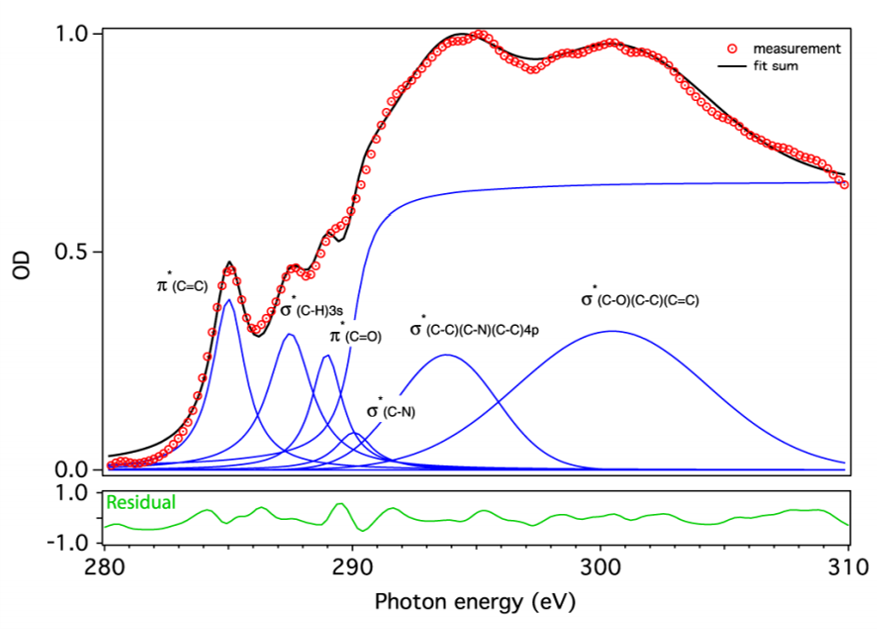
The photon flux of the HHG light source is the highest reported in the water window and, more important, it corresponds to the first isolated 355 attosecond duration soft x-ray pulse. Critical for absorption spectroscopy is the spectral stability, which is ensured through CEP stability of the laser source.3,4 The ICFO research group then demonstrated the utility of their tabletop HHG light source for performing carbon K-edge NEXAFS spectroscopy on a 200 nm freestanding polyimide film. Figure 4 shows the absorption spectrum that was taken in a mere 5 min with all peaks around the carbon K-edge clearly visible and identifiable from the known orbitals in polyimide.4
Figure 4: NEXAFS measurement of a 200 nm free-standing polyimide foil (red circles). A peak fit with known transitions (blue) agrees very well (black curve) with the measurement (4). A PIXIS-XO camera was used to acquire the data, which was extracted from a single 5 min integration. Courtesy of Prof. Dr. Jens Biegert (Institute of Photonic Sciences, Barcelona). First published in Opt. Lett. 39, 5383-5386 (2014).
This HHG instrument is the first reported high-flux tabletop source of coherent x-ray radiation in the water window that reaches a photon flux of (1.85 ± 0.12) x 107 photons/sec/1% bandwidth at 300 eV and the first isolated attosecond pulse in that regime.3,4
Enabling Technologies
The Princeton Instruments PIXIS-XO scientific camera utilized by the research team in Barcelona features a cooled, back-illuminated CCD without anti-reflective coating in order to facilitate the direct detection of ultra-low-energy x-rays. In addition to the camera’s software-selectable gains and readout speeds, a rotatable conflat flange with a high-vacuum-interface design makes the PIXIS-XO an excellent choice for ultra-high-vacuum applications.
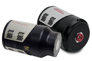
Princeton Instruments offers a variety of cameras for the direct detection of soft x-rays (see Figure 5), including not only the PIXIS-XO but the PIXIS-XB, which uses a thin beryllium window to vacuum seal the unit for deep cooling, protect its back-illuminated deep-depletion CCD, and reduce background by filtering low-energy x-rays.
Another Princeton Instruments scientific x-ray CCD camera, the PI-MTE, implements a thermoelectrically cooled design that utilizes PCBs thermally linked to circulating coolant in order to provide reliable operation inside vacuum chambers. The PI-MTE camera’s compact size and flexible tubing permit the positioning of the detector in limited space or on a movable arm.
Figure 5: PI-MTE, PIXIS-XO, and PIXIS-XO scientific x-ray CCD cameras from Princeton Instruments are designed to facilitate the direct detection of soft x-rays.
Future Trends
The successful use of a tabletop HHG instrument by researchers at ICFO to perform carbon K-edge NEXAFS spectroscopy within the water window embodies the trend towards miniaturization and personalization of coherent x-ray radiation sources for advanced x-ray techniques. The group’s most recently published HHG-enabled results, the spatio-temporal isolation of attosecond soft x-ray pulses in the water window,3 signifies yet another important stride on this march of technological progress.
Researchers from the Far East6 to the Far West2 are designing and utilizing a variety of tabletop HHG light sources to meet their own specific x-ray imaging and spectroscopy requirements. In addition to the obvious cost and access advantages over large-scale, state-of-the-art synchrotrons and XFEL sources, these compact HHG instruments are beginning to offer superior temporal resolution.3 Furthermore, HHG-enabled coherent x-ray diffraction imaging is able to produce quantitative, high-contrast phase and amplitude images in both transmission and reflection, as well as probe dynamic phenomena in three dimensions,2 offering a powerful complement to surface-scanning technologies such as atomic force microscopy.
Scientific x-ray CCD cameras, such as Princeton Instruments’ PIXIS-XO, PIXIS-XB, and PI-MTE, ensure the sensitivity, speed, and flexibility needed to match ongoing advances in tabletop HHG instrumentation. And as x-ray experimental setups for individual labs continue to evolve and gain popularity, selecting the right application-appropriate camera will be imperative to capitalize fully on the benefits of these new setups.
Adams D.E., Wood C.S., Murnane M.M., and Kapteyn H.C. Tabletop high harmonics illuminate the nano-world. LFW, 38–41 (2015).
Silva F., Teichmann S., Cousin S.L., Hemmer M., and Biegert J. Spatiotemporal isolation of attosecond soft x-ray pulses in the water window. Nat. Commun. 6 (2015).
Cousin S.L., Silva F., Teichmann S., Hemmer M., Buades B., and Biegert J. High-flux tabletop soft x-ray source driven by sub-2-cycle, CEP stable, 1.85-μm 1-kHz pulses for carbon K-edge spectroscopy. Opt. Lett. 39, 5383–5386 (2014).
Cousin S.L., Silva F., Teichmann S., Hemmer M., and Biegert J. Molecular fine structure from water window x-rays. OPN, 58 (2014).
Sung H., Kim Y.S., Cho W.J., Kim Y.T., Kim J.H., Lee S., and Jhon Y.M. Coherent EUV light source by high harmonic generation for EUV metrology. Recent Advances in Telecommunications and Circuits, 34–36, World Scientific and Engineering Academy and Society Press (2013). ISBN: 978-960-474-308-7
Coherent diffraction microscopy and its applications in nanoscience and biology. (accessed online Aug. 2015) http://www.physics.ucla.edu/research/imaging/Research/Research_1.pdf
Further Information
Related Products
Overview of the advantages of the products available which are optimized for soft x-ray microscopy applications.
High-Repetition Rate Laser-Matter Interactions for Solid TargetsCustomer Stories
Ted Biewer
Oak Ridge National Lab, Fusion Energy Division, Diagnostics and Control
Background
The fusion diagnostics and control group led by Ted Biewer at Oak Ridge National Laboratory specializes in measuring and monitoring properties of plasmas in fusion experiments. We talked to Drew Elliott, a scientist in the group: “What we do in our group is develop and operate diagnostics to better characterize a lot of these experiments.” While several groups and experiments worldwide are searching and developing ways to create more efficient and economical fusion experiments, measuring key parameters of fusion plasmas remains challenging. “We are diagnostic experts and are able to make more accurate measurements or make new measurements which other groups either don’t have the personnel, hardware or expertise.”
In an effort to bring their expertise to multiple fusion labs around the world, the researchers recently developed a compact and portable system that can be transported and operated at different experiment sites. The instrument implements a technique called Thomson scattering (scattering of electromagnetic radiation by charged particles) which Dr. Elliott explains as basically being a very accurate way to measure temperature or density in hot plasmas. Using optical methods is crucial in this process as physical probes would be destroyed in the hot and harsh environments found in fusion plasmas.
Using their new setup the group will be able to support experiments worldwide by giving them diagnostic capabilities and information that they did not have before.
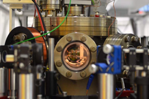
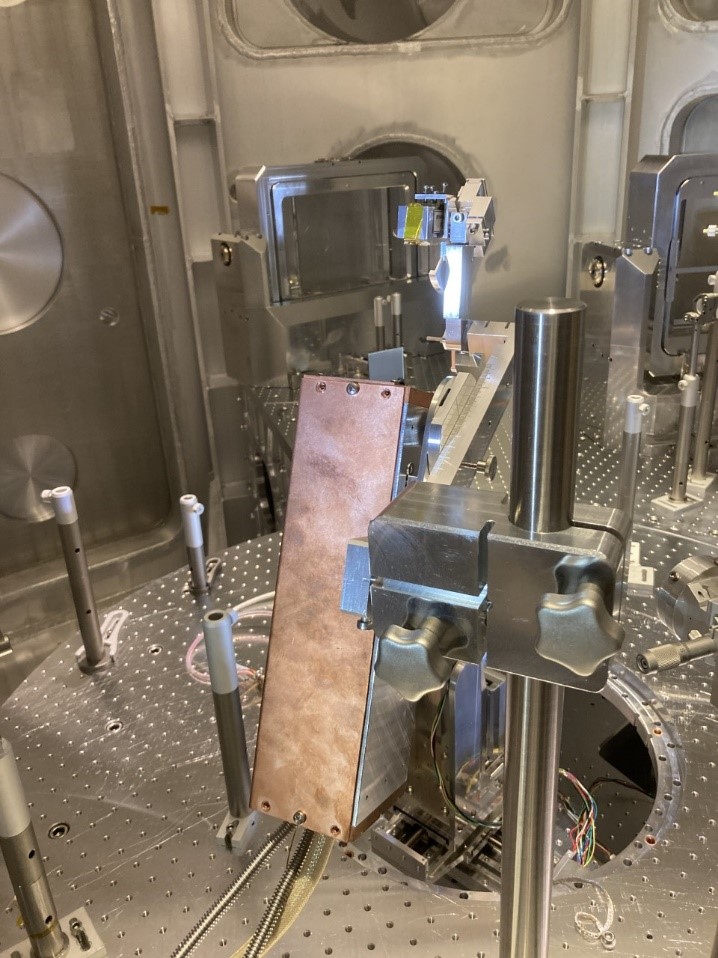
Figure 1: Image of the X-ray spectrometer in the vacuum chamber, with the PI-MTE inside its Faraday cage
Challenge
The main challenge in developing the new setup was that the spectroscopy system needed to be compact enough to be transportable, but also have flexibility to adapt to a wide range of experiments that could have very different parameters. For example, plasma temperatures might range from relatively cold plasmas (at around 1000 times room temperature) to temperatures approaching those inside of the sun. Dr. Elliott explained that the temperature limit that can be measured using their spectroscopic technique essentially depends on the spectral bandwidth of the spectrograph which should be able to adapt its spectral range for different experimental situations.
One problem, encountered in conventional spectrographs is that their instrument response function changes significantly between the center and edges of the camera sensor due to optical aberrations. Spectral lines would then change size and shape depending where on the sensor they are observed. The team required a better instrument response so complex and difficult corrections in data processing could be avoided.
Experiments on plasmas also require detectors capable of very short exposure times to suppress the very bright background radiation of the hot plasma. If the exposure is timed precisely with the signal from a short laser pulse, this background light can be effectively negligible compared to the laser signal.
“Overall, the IsoPlane and PI-MAX combination allowed us to set up a compact system that achieves the required measurement capability”
Solution
The group implemented a spectroscopy system consisting of IsoPlane-320 spectrographs and PI-MAX4 ICCD cameras for their transportable diagnostic system. The IsoPlane-320 has an advanced optical system, compared to conventional lab spectrographs, that minimizes optical aberrations. As a result, the instrument response and resolution remain constant across the detector. Low aberrations also allow for observation of a larger number of spectral tracks. Thomson scattering experiments often use multiple fiber optic inputs, addressing different locations in the experiment. This spectrometer and camera configuration allows the Oak Ridge team to observe all these channels simultaneously. IsoPlane spectrographs also come equipped with a triple grating turret, so the spectral bandwidth can be changed quickly by switching to gratings with larger or smaller groove density.
Moreover, the PI-MAX4 intensified CCD camera allows for nano-second gate times, necessary to suppress the bright plasma background. The gate opening can be synchronized with picosecond precision to the signal induced by the fast laser pulses.
“Overall, the IsoPlane and PI-MAX combination allowed us to set up a compact system that achieves the required measurement capability.”