
Introduction
Ultimately, the true worth of a scientific camera is determined by its ability to flexibly meet the performance requirements deemed most useful by a given researcher. As many disciplines have continued to evolve over recent years to encompass more varied investigatory techniques, sets of critical requirements have also expanded. When seeking a scientific camera to satisfy diverse needs, one must be sure to look for the most versatile, highest performance solutions available.
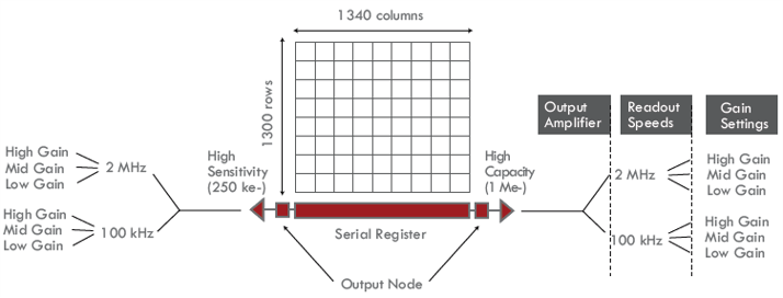
This note addresses some of the ways in which the flexible electronic architecture of Teledyne Princeton Instruments scientific CCD cameras extends application utility to facilitate research in a number of fields. The unique design of these CCD cameras features a pair of output amplifiers, each with its own software-selectable speed and gain settings, thereby enabling researchers to tailor performance to suit various x-ray, imaging, and spectroscopy applications.
Dual-Amplifier Design
Teledyne Princeton Instruments’ exclusive dual-amplifier configuration provides two independent amplifiers whose electronics are optimized for high-capacity readout and high-sensitivity readout, respectively (Fig.1). The high-capacity amplifier affords wider dynamic range by increasing full well capacity, in effect allowing each pixel of the CCD array to collect a greater number of photons. Generally speaking, the high-capacity amplifier is utilized for applications involving medium-energy x-rays or high levels of incident light. The high-sensitivity amplifier, meanwhile, is designed to deliver the lowest possible read noise at a given readout frequency. Typically, the high-sensitivity amplifier is utilized for applications involving low-energy x-rays or low levels of incident light.
Selectable Speed and Gain
Both the high-capacity and high-sensitivity amplifier offer their own independent, software-selectable speed and gain settings to allow researchers to fine-tune camera performance. Faster readout speeds can be used to acquire images quickly when the amount of incident light or energy is sufficient to do so without compromising results, or when setting up an experiment in focus mode. Slower readout speeds deliver high sensitivity by preserving dynamic range and boosting signal-to-noise ratio (SNR).
Note that signal-to-noise ratio refers to the relative magnitude of the signal compared to the uncertainty in that signal on a per-pixel basis. Specifically, it is the ratio of the measured signal to the overall measured noise (frame-to-frame) at that pixel. Accurate calculation of SNR must take into account the three primary sources of noise in a CCD camera: photon-flux-related shot noise, thermally generated dark noise, and the aforementioned read noise, as well as several additional noise sources. High SNR is particularly important in applications requiring precise light measurements.
Like selectable readout speeds, selectable gain settings also provide researchers a valuable tool for tailoring camera performance. Gain defines the number of electrons that correspond to a single analog-to-digital unit (e-/ADU). For example, a gain of 4 means the camera digitizes the CCD signal so that each ADU corresponds to 4 electrons. Prior to the application of gain, each photon detected by the CCD generates a given number of electrons based on the photon’s energy.
Soft X-ray Applications
Soft x-rays have photon energies approx. between 100 eV and 1000 eV (12.4 nm to 1.24 nm). These wavelengths allow researchers to utilize high-spatial-resolution x-ray microscopy techniques that offer good penetration in micrometer-thick specimens without the laborious sample preparation required by electron microscopy. Soft x-rays also enable elemental analysis of specimens by providing excellent intrinsic contrast between organic materials (e.g., proteins) and water in the so-called “water window” (284 eV to 543 eV) between the carbon and oxygen absorption edges.
At 250 eV, only 68.5 electrons are generated for each photon detected by the CCD; at 100 eV, just 27.4 electrons are generated per detected photon. Thus, when working with soft x-rays, the high-sensitivity amplifier should be utilized. Slower speed settings can further enhance sensitivity. In addition, the high-sensitivity amplifier’s gain settings should be used to optimize dynamic range and SNR to achieve the image contrast required. The x-ray energy itself can also be tuned to attain desired contrast.
Hard X-ray Applications
Hard x-rays have photon energies greater than 1000 eV (1 keV). One increasingly popular x-ray imaging application in this range is coherent x-ray diffraction imaging, also known as lensless x-ray diffraction imaging. In this technique, a CCD camera is used to detect the continuous diffraction pattern that results from shining a pink x-ray beam on a non-crystalline specimen. Use of coherent x-ray diffraction imaging is expanding from the soft x-ray range (for imaging biological specimens) into the 5 keV to 7 keV range (for imaging nanoparticles).
Another application that uses hard x-rays is x-ray photon correlation spectroscopy, or XPCS. In this technique, fluctuations in frequency and intensity of the scattered light (caused by temperature and pressure changes) in a sample are imaged at 8 keV.
Even at 1 keV, around the lower boundary of the medium-energy x-ray range, 274 electrons are generated for each photon detected by the CCD. Therefore, since additional sensitivity is not required to boost SNR, researchers should utilize the high-capacity amplifier when working with hard x-rays. Slower readout speeds and higher gain settings are typically less beneficial in this energy range due to the already sufficient sensitivity and ample signal.
Raman Spectroscopy
Raman spectroscopy is a highly popular technique for studying solids, liquids, and gases, is based on the scattering of monochromatic light. Excitation light is absorbed by a sample and then re-emitted; the resultant “Raman shift” provides information on vibrational, rotational, and other molecular modes. Since the pixel binning employed for this photon starved application increases noise as well as signal, the high-sensitivity amplifier should be used along with slower readout speeds and medium-to-high gain to boost SNR while preserving wide dynamic range.
Conclusion
The flexible electronic architecture of Teledyne Princeton Instruments scientific CCDs provides two distinct output amplifiers designed for optimum high-capacity and high-sensitivity readout, respectively. Camera performance can be further refined using each amplifier’s software-selectable speed and gain settings. This built-in versatility allows researchers to utilize an individual scientific camera for a greater range of applications without sacrificing high performance.
Further Information

QE Response Curves for the Soft X-Ray – VUV
Technical note describing how sensor design effects quantum efficiency for soft x-ray and VUV applications.
Direct Detection of X-rays using CCD Technology
The fundamentals of CCD technology and how it is optimized for the direct detection of x-rays.
Utilizing Fiberoptics for Indirect Detection of X-rays
Technical note describing the main specifications to consider when choosing fiberoptics for indirect detection of x-rays.